. 2011 Mar 9;6(3):e17838.
doi: 10.1371/journal.pone.0017838.

Renal thrombotic microangiopathy in mice with combined deletion of endocytic recycling regulators EHD3 and EHD4

Affiliations

Renal thrombotic microangiopathy in mice with combined deletion of endocytic recycling regulators EHD3 and EHD4

Manju George et al. PLoS One. .

Abstract

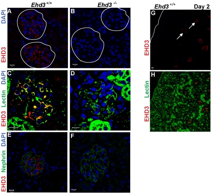
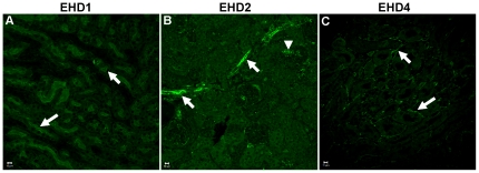
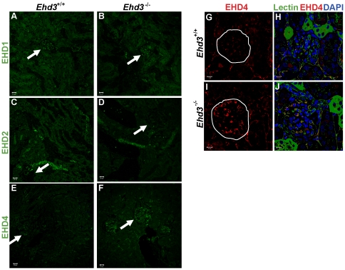
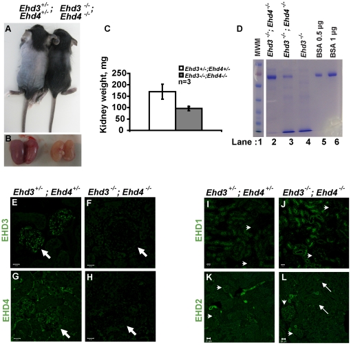
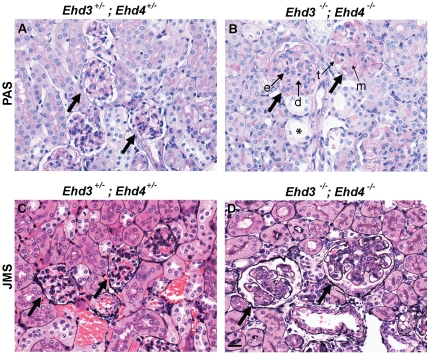
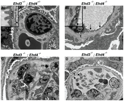
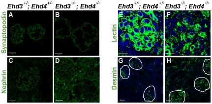
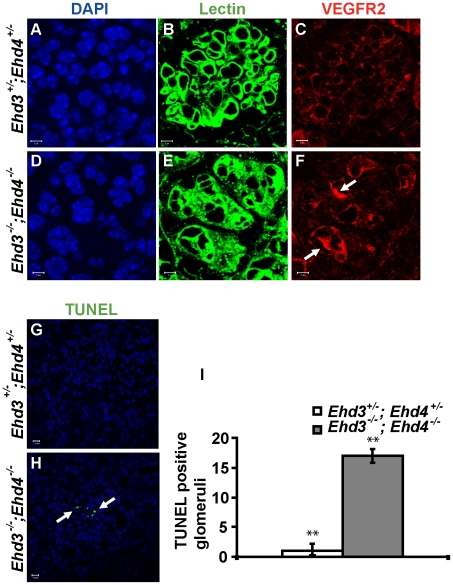
Eps15 Homology Domain-containing 3 (EHD3), a member of the EHD protein family that regulates endocytic recycling, is the first protein reported to be specifically expressed in the glomerular endothelium in the kidney; therefore we generated Ehd3(-/-) mice and assessed renal development and pathology. Ehd3(-/-) animals showed no overt defects, and exhibited no proteinuria or glomerular pathology. However, as the expression of EHD4, a related family member, was elevated in the glomerular endothelium of Ehd3(-/-) mice and suggested functional compensation, we generated and analyzed Ehd3(-/-); Ehd4(-/-) mice. These mice were smaller, possessed smaller and paler kidneys, were proteinuric and died between 3-24 weeks of age. Detailed analyses of Ehd3(-/-); Ehd4(-/-) kidneys demonstrated thrombotic microangiopathy (TMA)-like glomerular lesions including thickening and duplication of glomerular basement membrane, endothelial swelling and loss of fenestrations. Other changes included segmental podocyte foot process effacement, mesangial interposition, and abnormal podocytic and mesangial marker expression. The glomerular lesions observed were strikingly similar to those seen in human pre-eclampsia and mouse models of reduced VEGF expression. As altered glomerular endothelial VEGFR2 expression and localization and increased apoptosis was observed in the absence of EHD3 and EHD4, we propose that EHD-mediated endocytic traffic of key surface receptors such as VEGFR2 is essential for physiological control of glomerular function. Furthermore, Ehd3(-/-); Ehd4(-/-) mice provide a unique model to elucidate mechanisms of glomerular endothelial injury which is observed in a wide variety of human renal and extra-renal diseases.


Conflict of interest statement

Competing Interests: Ehd3 null mice and Ehd4 null mice were generated while MG, MAR, MN, GGY, RMG, CBG, VB and HB were at Evanston Northwestern Healthcare Research Institute, Evanston IL.

Figures

Figure 1
Figure 1. EHD3 expression in the glomerular endothelium.
(A–B) Three µm thick kidney sections from three month old male Ehd3+/+ and Ehd3–/– mice were stained using antibodies to EHD3 and confocal images acquired as described. EHD3 staining (red) and DAPI staining of nuclei (blue) are shown. White circles demarcate glomeruli. (C–D) Triple staining of kidney sections with labeled tomato lectin (green) and EHD3 (red) and DAPI (blue) shows colocalization between EHD3 and tomato lectin staining (yellow in panel C). (E–F) Triple staining with the podocyte marker, nephrin (green), EHD3 (red) and DAPI (blue) shows nephrin staining around endothelial EHD3 staining in panel E. EHD3 staining is absent in Ehd3–/– kidney sections as expected in panels B, D and F. (G–H) Kidney sections from 2 day old Ehd3+/+ mice were double stained with EHD3 (red) and lectin (green), yellow in panel H shows colocalization. The white line demarcates the kidney capsule in panel G and white arrows point to EHD3 expression in capillary loop stage glomeruli. S-shaped and comma shaped glomeruli that lack EHD3 staining are clearly visible in the nephrogenic zone in panel H. Scale bar = 10 µm.
Figure 2
Figure 2. Spatially distinct expression of EHD proteins in the kidney.
(A–C) Kidney sections from three month old male Ehd3+/+ and Ehd3–/– mice were processed and stained using antibodies to indicated EHD proteins and confocal images acquired as described in Materials and Methods. EHD staining is shown in green. Scale bar = 10 µm. White arrows point to EHD1 expression in brush border epithelium (panel A), EHD2 expression in the interlobular arteries (panel B), and EHD4 expression in the peritubular capillaries (panel C); the white arrow head points to EHD2 expression in afferent arterioles (panel B).
Figure 3
Figure 3. Compensatory increase in EHD4 in the Ehd3–/– glomerular endothelium.
(A–B) Confocal images of formalin-fixed, paraffin-embedded 3 µm thick kidney sections from 5 month old male Ehd3 +/+ and Ehd3–/– mice were immunostained with antibodies to EHD1, (C–D) EHD2 and (E–F) EHD4. EHD protein staining is shown in green. One glomerulus in each panel is denoted by a white arrow. Scale bar = 20 µm. (G–J) Sections triple stained for EHD4 (red, panel G–J), labeled tomato lectin (green) and DAPI (blue) are shown. Yellow in panel J shows colocalization between lectin and EHD4. White circles demarcate glomeruli. Scale bar = 10 µm.
Figure 4
Figure 4. Proteinuria in Ehd3 –/– ; Ehd4 –/– mice.
(A) Seventeen day-old Ehd3 –/– ; Ehd4 –/– and littermate Ehd3 +/– ; Ehd4 +/– mice were euthanized and photographed. (B) Kidneys dissected from mice shown in (A) were photographed, note the smaller and paler Ehd3 –/– ; Ehd4 –/– kidneys. (C) Kidneys weights from 3-week old Ehd3 –/– ; Ehd4 –/– mice and littermate controls were plotted, error bars indicate standard deviation. (D) Urine samples from 17 day old mice of indicated genotypes (lane 2 & 3) and a 3 month old Ehd3 –/– mouse (lane 4) were run on a 7.5% SDS-PAGE and stained with Coomassie Blue. Gels were scanned following de-staining. Bovine serum albumin (BSA) was used as a positive control (lanes 5 and 6). MWM, molecular weight marker (lane 1). Lanes 1–6 were run on the same gel but were noncontiguous. (E–H) 3 µm kidney sections from mice shown in (A) were stained for EHD3 and EHD4 (green) and confocal images acquired as described. White arrows point to expression of EHD3 and EHD4 in Ehd3 +/– ; Ehd4 +/– kidney sections (panel E and G) and absence of expression in Ehd3 –/– ; Ehd4 –/– kidney sections (panel F and H). (I–L) Kidney sections from mice shown in (A) were stained for EHD1 (green, panels I–J) and EHD2 (green, panels K–L) and confocal images acquired as described. White arrows point to expression of EHD1 and EHD2 in kidney sections, while white arrow heads point to glomeruli. Scale bar = 20 µm.
Figure 5
Figure 5. Ehd3–/–; Ehd4–/– mice develop thrombotic microangiopathy.
(A–B) Kidney sections from 22 day old Ehd3–/–; Ehd4–/– mice and littermate controls were stained with PAS to visualize tubular and glomerular basement membranes. Healthy capillary loops are seen in the Ehd3 +/–; Ehd4 +/– glomeruli (panel A) while Ehd3–/–; Ehd4–/– kidney sections showed endothelial swelling (e), enlarged glomeruli with segmental thickening (t), duplication of basement membranes (d) and mesangiolysis (m). Some tubular dilation is also seen (asterisks). (C–D) Jones methenamine silver (JMS) staining of kidney sections from Ehd3–/–; Ehd4–/– mice (panel D) and littermate controls (panel C) show duplicated and thickened glomerular basement membrane, expanded mesangium and relatively avascular glomeruli in Ehd3–/–; Ehd4–/– kidney sections. Thick black arrows point to glomeruli.
Figure 6
Figure 6. Ultrastructural changes in Ehd3–/–; Ehd4–/– glomeruli.
(A) Endothelial cells with fenestrations (black arrow heads) and podocytes with intact foot processes (white arrows) are evident in the Ehd3 +/–; Ehd4 +/– section. (Ai) Inset shows a representative higher magnification image, inset scale bar = 500 nm. (B) Abnormally thickened and lamellated basement membranes (t), swollen endothelium without fenestrations and effacement of podocytes foot processes were seen in the Ehd3–/–; Ehd4–/– kidney section. Multiple small “holes” presumed to be abnormal endosomes were seen in the endothelium (black arrows); these structures are clear in the higher magnification image in B. Inset scale bar = 500 nm. Scale bars = 2 µm. (C–D) Images from Ehd3–/–; Ehd4–/– kidneys also show additional changes including mesangiolysis (m), mesangial interposition (i), flocculent subendothelial material (f) and platelet (p) accumulation in a capillary loop. GBM, glomerular basement membrane. Scale bars = 5 µm.
Figure 7
Figure 7. Alteration in endothelial, podocytic and mesangial cells in Ehd3–/–; Ehd4–/– glomeruli.
(A–D, G–H) Kidney sections from 39 day old Ehd3 +/–; Ehd4 +/– and Ehd3–/–; Ehd4–/– mice were immunostained with antibodies to synaptopodin (A–B), nephrin (C–D) and desmin (G–H) as described in Materials and Methods. (E–F) Endothelial cells were visualized by labeled tomato lectin staining (green) and DAPI (blue) was used to counterstain nuclei. Markedly altered staining patterns were observed in the Ehd3–/–; Ehd4–/– kidney sections in each case. Scale bar = 20 µm in panels A–F and 10 µm in panels G–H. White lines demarcate glomeruli in panels G–H.
Figure 8
Figure 8. Altered VEGFR2 localization and increased apoptosis in Ehd3–/–; Ehd4–/– glomeruli.
(A–F) Kidney sections from Ehd3+/–; Ehd4+/– and Ehd3–/–; Ehd4–/– mice were immunostained with antibodies to VEGFR2 (red). Labeled lectin (green) was used to mark endothelial cells (B and E), while DAPI (blue) stained nuclei (A and D). Uniform low level of VEGFR2 staining in seen in lectin positive glomerular endothelial cells in the Ehd3+/–; Ehd4+/– glomeruli (panel C), while diffuse and abnormal lectin staining was seen in the Ehd3–/–; Ehd4–/– glomeruli (panel F), few lectin positive cells showed intense VEGFR2 staining (white arrows, panel F). (G–I) Kidney sections from Ehd3+/–; Ehd4+/– and Ehd3–/–; Ehd4–/– mice were subjected to TUNEL assay as described in Materials and Methods. Confocal images of TUNEL staining (green) are shown (panels G–H), white arrow points to apoptotic nuclei (panel H). About 45 glomeruli each were counted in 12 kidney sections to arrive at the graph (panel I). Error bars indicate standard deviation (**indicates P<0.05 using two-tailed analysis).

References

    1. Ballermann BJ. Contribution of the endothelium to the glomerular permselectivity barrier in health and disease. Nephron Physiol. 2007;106:p19–25. - PubMed
    1. Mundel P, Reiser J, Zuniga Mejia Borja A, Pavenstadt H, Davidson GR, et al. Rearrangements of the cytoskeleton and cell contacts induce process formation during differentiation of conditionally immortalized mouse podocyte cell lines. Exp Cell Res. 1997;236:248–258. - PubMed
    1. Abrahamson DR. Development of kidney glomerular endothelial cells and their role in basement membrane assembly. Organogenesis. 2009;5:275–287. - PMC - PubMed
    1. Haraldsson B, Nystrom J, Deen WM. Properties of the glomerular barrier and mechanisms of proteinuria. Physiol Rev. 2008;88:451–487. - PubMed
    1. Patrakka J, Tryggvason K. Molecular make-up of the glomerular filtration barrier. Biochem Biophys Res Commun. 2010;396:164–169. - PubMed

Publication types

MeSH terms

LinkOut - more resources