. 2011 Mar 9;6(3):e17850.
doi: 10.1371/journal.pone.0017850.

Anti-tumor activity of a novel compound-CDF is mediated by regulating miR-21, miR-200, and PTEN in pancreatic cancer

Affiliations

Anti-tumor activity of a novel compound-CDF is mediated by regulating miR-21, miR-200, and PTEN in pancreatic cancer

Bin Bao et al. PLoS One. .

Retraction in

Abstract

Background: The existence of cancer stem cells (CSCs) or cancer stem-like cells in a tumor mass is believed to be responsible for tumor recurrence because of their intrinsic and extrinsic drug-resistance characteristics. Therefore, targeted killing of CSCs would be a newer strategy for the prevention of tumor recurrence and/or treatment by overcoming drug-resistance. We have developed a novel synthetic compound-CDF, which showed greater bioavailability in animal tissues such as pancreas, and also induced cell growth inhibition and apoptosis, which was mediated by inactivation of NF-κB, COX-2, and VEGF in pancreatic cancer (PC) cells.

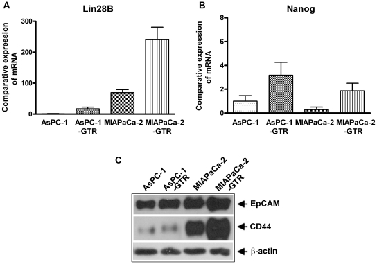
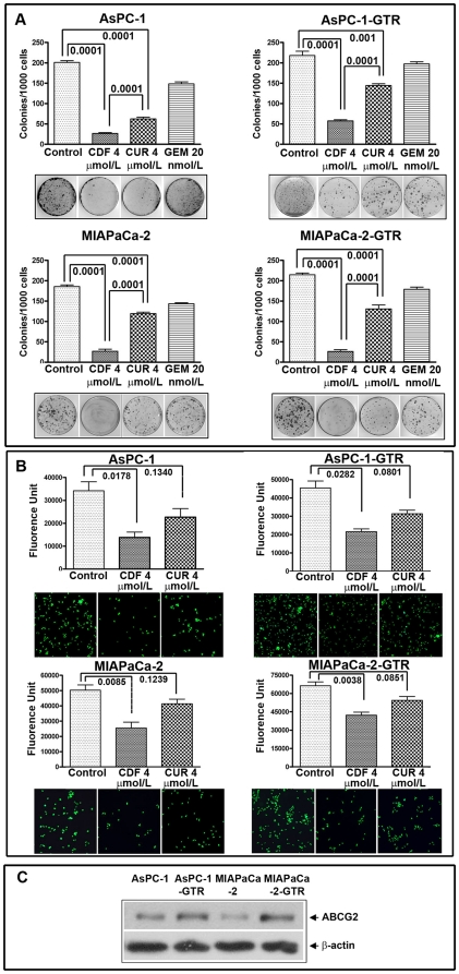
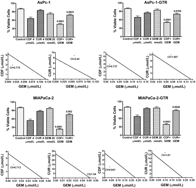
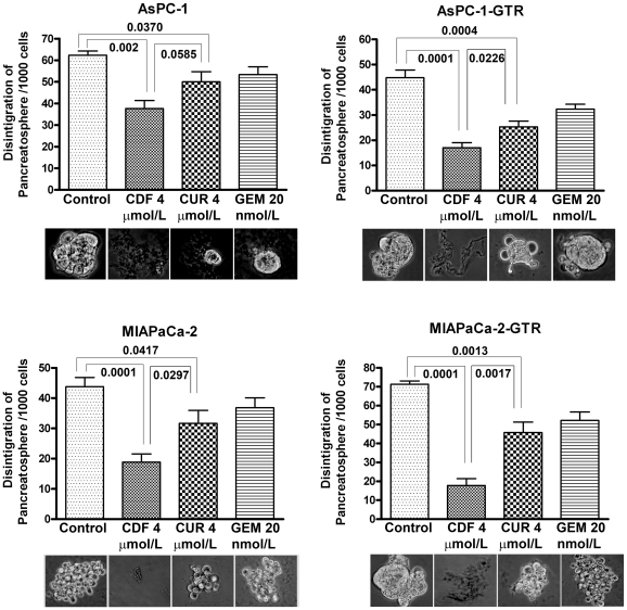
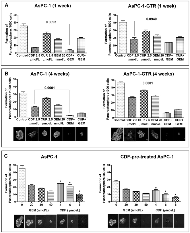
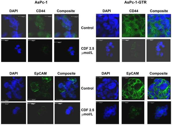
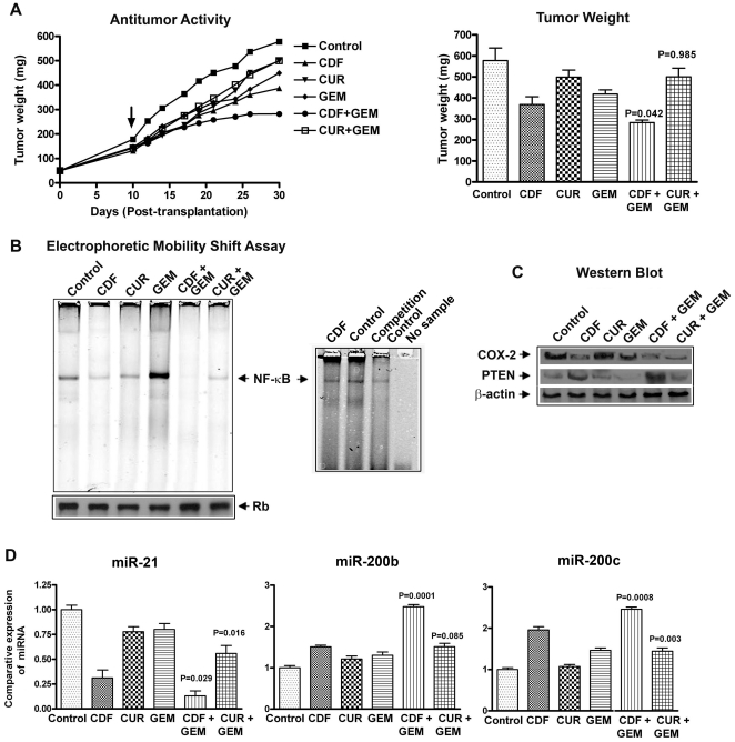
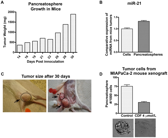
Methodology/principal findings: In the current study we showed, for the first time, that CDF could significantly inhibit the sphere-forming ability (pancreatospheres) of PC cells consistent with increased disintegration of pancreatospheres, which was associated with attenuation of CSC markers (CD44 and EpCAM), especially in gemcitabine-resistant (MIAPaCa-2) PC cells containing high proportion of CSCs consistent with increased miR-21 and decreased miR-200. In a xenograft mouse model of human PC, CDF treatment significantly inhibited tumor growth, which was associated with decreased NF-κB DNA binding activity, COX-2, and miR-21 expression, and increased PTEN and miR-200 expression in tumor remnants.

Conclusions/significance: These results strongly suggest that the anti-tumor activity of CDF is associated with inhibition of CSC function via down-regulation of CSC-associated signaling pathways. Therefore, CDF could be useful for the prevention of tumor recurrence and/or treatment of PC with better treatment outcome in the future.


Conflict of interest statement

Competing Interests: The authors have declared that no competing interests exist.

Figures

Figure 1
Figure 1. Comparative expression of Lin28B (A) and Nanog (B) mRNA by qRT-PCR showed increased expression in resistant cell lines compared to parental cell lines, supporting the CSC characteristics of these cell lines.
The characteristics of CSCs were further confirmed by the protein expression of EpCAM and CD44 (C).
Figure 2
Figure 2. CDF and Curcumin decreased clonogenicity and invasion in AsPC-1, AsPC1-GTR, MIAPaCa-2, and MIAPaCa-2-GTR cells. Clonogenic assay (A) Invasion assay (B).
Fluorescence of the invaded cells was read using ULTRA Multifunctional microplate reader (TECAN) at excitation/emission wavelengths of 530/590 nm. Basal level of ABCG2 expression showing relatively higher expression in drug resistant cell lines (C).
Figure 3
Figure 3. CDF and its combination with gemcitabine inhibited cell viability.
MTT assay was conducted in all four cell lines after 72h of treatment with CDF, curcumin, or its combination with gemcitabine. Untreated control has been assigned a value of 100%. The p value shown represents comparisons between single agent and their combinations by using paired t-test. The combination Index (CI) <1 for CDF and Gemcitabine combination indicates synergism.
Figure 4
Figure 4. CDF remarkably increased disintegration of pancreatospheres in AsPC-1, AsPC1-GTR, MIAPaCa-2, and MIAPaCa-2-GTR cells.
P values were calculated by the paired t test.
Figure 5
Figure 5. CDF decreased the formation of pancreatospheres in AsPC-1 and AsPC-1-GTR cells 1 week treatment (A); 4 weeks treatment (B); AsPC-1 and CDF-pre-treated AsPC-1 cells treated with gemcitabine and CDF (C).
A significant reduction in pancreatospheres was observed in cells treated with CDF shown by asterisk
Figure 6
Figure 6. CDF treatment decreased the expression of CD44 and EpCAM, the known markers of CSCs.
Expression in pancreatospheres of AsPC-1 and AsPC-1-GTR cells was assessed by confocal microscopy (Magnification X250).
Figure 7
Figure 7. CDF exhibited anti-tumor activity in MIAPaCa-2 cells induced tumors in a xenograft mouse model, which was consistent with inhibition of NF-κB DNA binding, COX-2, miR-21, and caused re-expression of miR200 in tumor remnants.
Anti-tumor activity and changes in tumor weight from each group of animals (A). The arrow indicates starting day of the treatment. NF-κB DNA binding activity of tumor tissues; and NF-κB competition control study with unlabeled NF-κB oligonucleotide (B). Western blots analysis of COX-2, PTEN and β-actin expression in tumor remnants (C); miR-21, miR-200b and miR-200c expression in tumor remnants as measured by real-time RT-PCR (D). P values were calculated by the paired t test.
Figure 8
Figure 8. Tumor growth pattern of pancreatospheres derived from MIAPaCa-2 cells.
(A). 5,000 pancreatospheres were inoculated in mice using 1∶1 matrigel, progressive tumor growth over a period of 30 days. Moderate increase in the expression of miR-21 as measured by real-time RT-PCR was observed in tumors derived from pancreatospheres compared to tumors derived from parental cells by injecting one million cells and tumor was assessed over the same period of time (B). Photographs showing tumor growth, arrow points to tumor and asterisk (*) refers to loco-regional lymph node metastasis whereas we did not find any metastasis when one million parental cells were injected (C). Tumor cells harvested from the tumors derived from pancreatospheres were treated with CDF showed significant inhibition in the formation of pancreatospheres (D).

References

    1. Jemal A, Siegel R, Ward E, Hao Y, Xu J, et al. Cancer statistics, 2009. CA Cancer J Clin. 2009;59:225–249. - PubMed
    1. Gaviraghi M, Tunici P, Valensin S, Rossi M, Giordano C, et al. Pancreatic cancer spheres are more than just aggregates of stem marker positive cells. Biosci Rep 2010 - PMC - PubMed
    1. Hermann PC, Bhaskar S, Cioffi M, Heeschen C. Cancer stem cells in solid tumors. Semin Cancer Biol. 2010;20:77–84. - PubMed
    1. Ischenko I, Seeliger H, Kleespies A, Angele MK, Eichhorn ME, et al. Pancreatic cancer stem cells: new understanding of tumorigenesis, clinical implications. Langenbecks Arch Surg. 2010;395:1–10. - PubMed
    1. Lee CJ, Dosch J, Simeone DM. Pancreatic cancer stem cells. J Clin Oncol. 2008;26:2806–2812. - PubMed

Publication types

MeSH terms