. 2011 Mar 9;6(3):e17901.
doi: 10.1371/journal.pone.0017901.

Changes in the organization of excitation-contraction coupling structures in failing human heart

Affiliations

Changes in the organization of excitation-contraction coupling structures in failing human heart

David J Crossman et al. PLoS One. .

Erratum in

  • PLoS One. 2011;6(4). doi:10.1371/annotation/061613ea-0f01-420f-bc3f-af36e5c35790. Ruygrok, Peter R [corrected to Ruygrok, Peter N]

Abstract

Background: The cardiac myocyte t-tubular system ensures rapid, uniform cell activation and several experimental lines of evidence suggest changes in the t-tubular system and associated excitation-contraction coupling proteins may occur in heart failure.

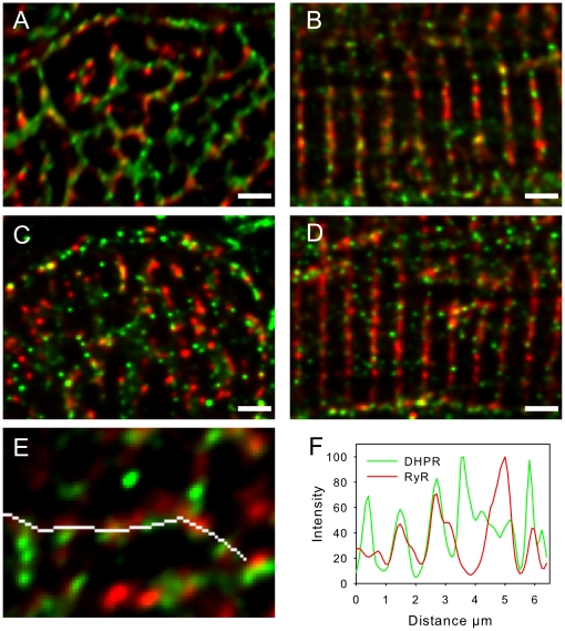
Methods and results: The organization of t-tubules, L-type calcium channels (DHPRs), ryanodine receptors (RyRs) and contractile machinery were examined in fixed ventricular tissue samples from both normal and failing hearts (idiopathic (non-ischemic) dilated cardiomyopathy) using high resolution fluorescent imaging. Wheat germ agglutinin (WGA), Na-Ca exchanger, DHPR and caveolin-3 labels revealed a shift from a predominantly transverse orientation to oblique and axial directions in failing myocytes. In failure, dilation of peripheral t-tubules occurred and a change in the extent of protein glycosylation was evident. There was no change in the fractional area occupied by myofilaments (labeled with phalloidin) but there was a small reduction in the number of RyR clusters per unit area. The general relationship between DHPRs and RyR was not changed and RyR labeling overlapped with 51±3% of DHPR labeling in normal hearts. In longitudinal (but not transverse) sections there was an ∼30% reduction in the degree of colocalization between DHPRs and RyRs as measured by Pearson's correlation coefficient in failing hearts.

Conclusions: The results show that extensive remodelling of the t-tubular network and associated excitation-contraction coupling proteins occurs in failing human heart. These changes may contribute to abnormal calcium handling in heart failure. The general organization of the t-system and changes observed in failure samples have subtle differences to some animal models although the general direction of changes are generally similar.


Conflict of interest statement

Competing Interests: The authors have declared that no competing interests exist.

Figures

Figure 1
Figure 1. Panel A shows tagged MRI short axis image through the middle of a failing heart in end diastole.
Tracked grids are shown as yellow overlay with sampled region marked in red. Panel B shows circumferential shortening (CC%) of the region indicated in the panel A compared to a similar region in normal human heart. Panel C shows RyRs (green) and WGA (red) labeling of diseased myocytes from the region shown in panel A. Image is z projections of 10 slices with z depth of 2.5 µm from deconvolved image stack. Scale bar is 10 µm.
Figure 2
Figure 2. WGA labelling of t-tubules in normal and failing human ventricular myocytes.
The top row shows images from normal cells in longitudinal and transverse sections (left to right) and corresponding images from diseased tissue is shown in the lower two rows. (A) Longitudinal sections of normal tissue shows uniformly spaced t-tubules. Occasional axial elements can also be seen. (B) A magnified view of the region shown by the box in A. (C) Normal myocyte in transverse section. A radial “spoke-like” organization of t-tubules is apparent. (D) Enlarged view of the region shown by the box in C. (E, I, K) Longitudinal sections from three different diseased cells, demonstrating the range of t-tubular morphologies found in disease with corresponding (F, J, L) magnified views. Note that while the enlarged view in L appears relatively normal, other regions with the same cell (K) are clearly abnormal. (G) Transverse section showing that, while the general direction of diseased tubules is radial, tubules are more disorganized. (H) Magnified view of the region shown by the box in G. Images are projections of 5 slices with z depth of 1 µm. Scale bars in overview images are 10 µm and in close up images 2 µm.
Figure 3
Figure 3. Analysis of changes in ventricular myocyte t-tubule geometry in human heart failure.
Correlation analysis of t-tubule angle: To illustrate the analysis, two model t-tubule images were created and correlated to the reference image, one with tubules running at 90° angle (panel A) and the other with tubules running at 45° angle (panel B), demonstrating that the angle with peak correlation intensity matches the direction of tubules in the model images. (C) Correlation analysis of longitudinal sections to measure t-tubule orientation referred to the cell surface. Analyses of normal cells are shown in the left panel and diseased tissue are shown on the right. Note the change in predominant tubule angle and increased spread of t-tubule angles in the diseased tissue. (D) Full width at half maximum (FWHM) measurements of control and diseased tubules corrected for optical blurring. In normal myocytes, tubule widths are smaller and have a narrower size distribution compared to diseased myocytes (p = 0.0003 see text).
Figure 4
Figure 4. Sarcolemmal protein labelling with WGA in normal and failing human cardiac myocytes.
Panels are arranged so that the left column corresponds to normal and the right column diseased tissue. A and B show longitudinal sections of control and diseased myocytes respectively, labelled for DHPRs (green) and WGA (red). The DHPR labelling is more continuous across the cell than the WGA label. Panels C and D show transverse images labelled for NCX (green) and WGA (red). In control myocytes (C), finer tubules containing only the NCX label can be seen to connect to larger spoke like tubules containing both labels. Diseased (D) myocytes have similar labelling pattern but many connections between larger tubules appear broken. Panels E and F show transverse sections labelled for Cav3 (green) and WGA (red). (E) Again, finer tubules labelled with only Cav3 connect larger tubules containing both labels. Diseased myocytes (F) have similar labelling pattern but with a reduction in fine Cav3 labelling between larger tubules. Images are z projections of 4 slices with z depth of 0.8 µm from deconvolved image stacks. Scale bars are 2 µm.
Figure 5
Figure 5. Comparison of myofilaments, t-tubules and RyR localization.
Panels A and B show transverse sections of normal and diseased myocytes respectively, labelled for Cav3 and f-actin. The contractile machinery is surrounded by Cav3 labelling at this plane which was selected to be centred at the z-line. Gaps in the centre of contractile bundles contain isolated Cav3 labelling (small white arrow) and inspection of adjacent sections show that this arises from axial tubules that join transverse tubules outside the presented image plane. A connection between adjacent radial t-tubules is indicated by the large arrow. Panels C and D show transverse sections of normal and diseased tissue (respectively) labelled for RyR and f-actin. It is apparent that each contractile bundle is surrounded by several RyR clusters. All images are projections of 4 slices with z depth of 0.8 µm. Panel E shows analysis of cell volume occupied by f-actin and no significant difference (P = 0.81) exists between normal and diseased tissue. Panel F shows analysis of the number of RyR clusters per µm3 of f-actin and there was a significant reduction in the density of RyR clusters in disease cells (*P = 0.03). Scale bars are 2 µm.
Figure 6
Figure 6. RyR and DHPR cluster colocalization is reduced in failing human cardiac myocytes.
Panels A and B show images of control myocytes in transverse and longitudinal orientation respectively, labelled for RyR (red) and DHPR (green). Panels C and D show images of diseased myocytes in transverse and longitudinal orientation respectively, labelled for RyR (red) and DHPR (green). Images are z projections of 4 optical sections with z depth of 1 µm. Scale bars are 2 µm. Panel E shows an exemplar region of DHPR and RyR labelling in a normal cell, this image is a single optical section. The white line indicates the position of intensity readings along a WGA labelled t-tubule (label not shown for clarity). Panel F shows the intensity profile of DHPR and RyR labelling along the white line shown in panel E. Similar results were seen with an alternative DHPR antibody (see methods and supplementary Figure S1).

References

    1. Bers DM. Cardiac excitation-contraction coupling. Nature. 2002;415:198–205. - PubMed
    1. Cannell MB, Cheng H, Lederer WJ. Spatial non-uniformities in [Ca2+]i during excitation-contraction coupling in cardiac myocytes. Biophys J. 1994;67:1942–1956. - PMC - PubMed
    1. Cannell MB, Soeller C. Sparks of interest in cardiac excitation-contraction coupling. Trends Pharmacol Sci. 1998;19:16–20. - PubMed
    1. Franzini-Armstrong C. Functional implications of RyR-dHPR relationships in skeletal and cardiac muscles. Biol Res. 2004;37:507–512. - PubMed
    1. Cheng H, Cannell MB, Lederer WJ. Propagation of excitation-contraction coupling into ventricular myocytes. Pflugers Arch. 1994;428:415–417. - PubMed

Publication types

MeSH terms

Substances