. 2011 Mar 8;6(3):e17649.
doi: 10.1371/journal.pone.0017649.

A novel extracellular Hsp90 mediated co-receptor function for LRP1 regulates EphA2 dependent glioblastoma cell invasion

Affiliations

A novel extracellular Hsp90 mediated co-receptor function for LRP1 regulates EphA2 dependent glioblastoma cell invasion

Udhayakumar Gopal et al. PLoS One. .

Abstract

Background: Extracellular Hsp90 protein (eHsp90) potentiates cancer cell motility and invasion through a poorly understood mechanism involving ligand mediated function with its cognate receptor LRP1. Glioblastoma multiforme (GBM) represents one of the most aggressive and lethal brain cancers. The receptor tyrosine kinase EphA2 is overexpressed in the majority of GBM specimens and is a critical mediator of GBM invasiveness through its AKT dependent activation of EphA2 at S897 (P-EphA2(S897)). We explored whether eHsp90 may confer invasive properties to GBM via regulation of EphA2 mediated signaling.

Principal findings: We find that eHsp90 signaling is essential for sustaining AKT activation, P-EphA2(S897), lamellipodia formation, and concomitant GBM cell motility and invasion. Furthermore, eHsp90 promotes the recruitment of LRP1 to EphA2 in an AKT dependent manner. A finding supported by biochemical methodology and the dual expression of LRP1 and P-EphA2(S897) in primary and recurrent GBM tumor specimens. Moreover, hypoxia mediated facilitation of GBM motility and invasion is dependent upon eHsp90-LRP1 signaling. Hypoxia dramatically elevated surface expression of both eHsp90 and LRP1, concomitant with eHsp90 dependent activation of src, AKT, and EphA2.

Significance: We herein demonstrate a novel crosstalk mechanism involving eHsp90-LRP1 dependent regulation of EphA2 function. We highlight a dual role for eHsp90 in transducing signaling via LRP1, and in facilitating LRP1 co-receptor function for EphA2. Taken together, our results demonstrate activation of the eHsp90-LRP1 signaling axis as an obligate step in the initiation and maintenance of AKT signaling and EphA2 activation, thereby implicating this pathway as an integral component contributing to the aggressive nature of GBM.


Conflict of interest statement

Competing Interests: The authors have declared that no competing interests exist.

Figures

Figure 1
Figure 1. eHsp90-LRP1 regulates EphA2 dependent motility and invasion in GBM.
Interference with eHsp90-LRP1 signaling inhibits GBM cell motility and invasion. (A, B) Treatment of cells with NPGA (1 µM) or anti-Hsp90 antibodies (20 ug/ml) (A), or suppression of LRP1 (B) similarly impaired G48a cell motility in wound healing assays. NS shRNA represents a nonspecifc shRNA control sequence. Percent migration is normalized to the 16 hr control and values represent the mean ± SD from 3 independent experiments (*p<0.001). (C) Serum starved parental or LRP1 silenced cells were added to top chambers of a Boyden assay and serum induced chemotaxis initiated in the presence of vehicle or NPGA. Cell numbers represent the mean ± SD from five random fields (*p<0.001). (D) The effects of eHsp90 targeting upon cell invasion were assessed by a Matrigel assay in the presence or absence of NPGA. Data is represented as the mean (± SD) of three replicates. *p<0.001. (E–F) Interference with eHsp90 function does not further inhibit cell motility or invasion in tandem with EphA2 silencing. G48a cells were transduced with either nonspecific (NS shRNA) or shEphA2 and effects of NPGA upon cell motility assessed in Boyden (right panel), and invasion assessed by Matrigel (left panel).
Figure 2
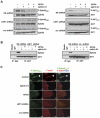
Figure 2. eHsp90-LRP1 signaling regulates activated EphA2 (phospho-S897), its association with AKT, and lamellipodia formation.
(A) Control or LRP1 silenced G48a cells were treated with NPGA, ephrin A1, or the combination, and the effects upon P-AKTS473 and P-EphA2S897 were evaluated. The effect of src silencing was included for relative comparison. (B) Interference with eHsp90 signaling (NPGA or LRP1 silencing), or AKT activation (src silencing or treatment with ephrin A1) disrupts EphA2-AKT protein complexes. (C) G48a cells grown were fixed 4 hr after wounding, followed by immunostaining with the indicated antibodies. Arrows indicate the leading edge localization of P-EphA2S897 (a–c). Stimulation of cells with ephrinA1 was included as positive control for suppression of EphA2S897 phosphorylation (d–f). Lamellipodia formation and concomitant localization of P-EphA2S897 is similarly suppressed by NPGA (g–i) or LRP1 silencing (j–l), or by src silencing (m–o). Scale bars 25 µm.
Figure 3
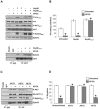
Figure 3. Preservation of AKT activation is required for lamellipodia formation, and concomitant cell motility and invasion.
(A) Serum starved (8 hr) G48a cells were exposed to either native or Hsp90ΔATPase proteins (3 µg/ml) for 15 min in the presence or absence of NPGA, and the indicated signaling molecules evaluated by immunoblot. The effect of these treatments upon EphA2-AKT interaction was also evaluated. (B) A Matrigel invasion assay was utilized to evaluate the ability of native or Hsp90ΔATPase proteins to sustain cell invasion in the presence of NPGA. Values represent the mean (± SD) of 3 independent experiments. (C) G48a cells stably transduced with the indicated HA-tagged myristolyated AKT constructs were exposed to NPGA and HA immunopurified complexes were evaluated for P-AKT and P-EphA2S897. (D) The pro-motility function of the indicated AKT proteins was evaluated in the presence or absence of NPGA using a scratch wound assay.
Figure 4
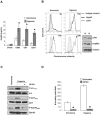
Figure 4. Hypoxic conditions amplify eHsp90-LRP1 initiated AKT-EphA2 signaling.
(A) The ability of hypoxia to modulate Hsp90α secretion was determined by ELISA, as in Supplementary Figure S1E. (B) G48a cells cultured in 1% serum were exposed to hypoxia (1% O2) for 36 hr and surface expression of Hsp90α and LRP1 was determined in intact cells by flow cytometry. A corresponding immunoblot shows total cellular expression of LRP1 and Hsp90α. (C) Representative immunoblot demonstrating effects of hypoxia upon activation of src, AKT, and EphA2 in the presence or absence of NPGA. (D) A Matrigel invasion assay was utilized to evaluate the effects of hypoxia (16 hr) upon cell invasion in the presence or absence of NPGA.
Figure 5
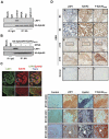
Figure 5. LRP1 is a co-receptor for EphA2 and co-localizes with P-EphA2S897 in clinical GBM specimens.
(A) HA-EphA2 transfected U87 cells were treated with NPGA, ephrin A1, or the src inhibitor PP2 for 16 hr. LRP1 was detected from HA immunoprecipitates. (B) U87 cells were transfected with HA-tagged wild type or point mutant (S897G) EphA2 plasmid, and LRP1 was detected from HA immunoprecipitates. (C) U87 cells were immunostained with indicated antibodies showing that LRP1 co-localized with EphA2. Scale bar 25 µm. The bottom panels represent magnified areas of confocal images derived from the respective upper panels, as delineated by the boxed region. Scale bar 5 µm. (D) Detection of P-EphA2S897 and LRP1 in human GBM specimens. Panels a–f, and m–o are paraffin sections, panels g–l are frozen specimens. Magnification ×200. Lower panels (a–l) are paraffin sections from the recurrent GBM specimens. Magnification ×400.
Figure 6
Figure 6. Molecular crosstalk between eHSP90-LRP1 and AKT-EphA2 signaling.
An eHSP90/LRP1 signaling axis is required to sustain src directed AKT activation, AKT dependent P-EphA2S897, and LRP1 recruitment to EphA2. These signaling events facilitate lamellipodia formation and support GBM cell motility and invasion. Hypoxia amplifies eHsp90 signaling and corresponding motility via enhanced LRP1 expression and Hsp90 secretion. NPGA inhibits eHsp90 signaling, with consequent inhibition of AKT, disruption of EphA2 and LRP1 complexes, and blockade of cell motility. ephrin A1 ligand similarly suppresses AKT activation, P-EphA2S897, EphA2-LRP1 complexes, and elicits comparable inhibitory effects upon GBM cell motility and invasion.

References

    1. Stupp R, Hegi ME, Mason WP, van den Bent MJ, Taphoorn MJ, et al. Effects of radiotherapy with concomitant and adjuvant temozolomide versus radiotherapy alone on survival in glioblastoma in a randomised phase III study: 5-year analysis of the EORTC-NCIC trial. Lancet Oncol. 2009;10:459–466. - PubMed
    1. Drappatz J, Norden AD, Wen PY. Therapeutic strategies for inhibiting invasion in glioblastoma. Expert Rev Neurother. 2009;9:519–534. - PubMed
    1. Glas M, Rath BH, Simon M, Reinartz R, Schramme A, et al. Residual tumor cells are unique cellular targets in glioblastoma. Annal Neurol. 2010;68:264–269. - PMC - PubMed
    1. Molina JR, Hayashi Y, Stephens C, Georgescu MM. Invasive glioblastoma cells acquire stemness and increased Akt activation. Neoplasia. 2010;12:453–463. 2887086. - PMC - PubMed
    1. Wykosky J, Debinski W. The EphA2 receptor and ephrinA1 ligand in solid tumors: function and therapeutic targeting. Mol Cancer Res. 2008;6:1795–1806. - PMC - PubMed

Publication types

MeSH terms