. 2011 Mar 8;6(3):e16746.
doi: 10.1371/journal.pone.0016746.

Mutations in PINK1 and Parkin impair ubiquitination of Mitofusins in human fibroblasts

Affiliations

Mutations in PINK1 and Parkin impair ubiquitination of Mitofusins in human fibroblasts

Aleksandar Rakovic et al. PLoS One. .

Abstract

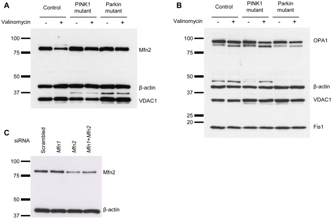
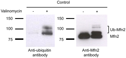
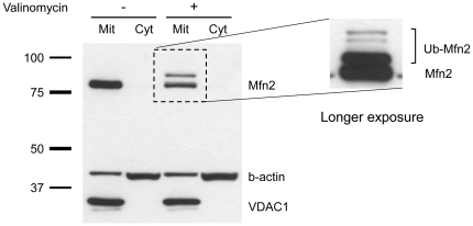
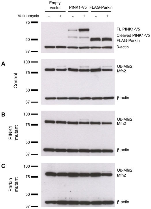
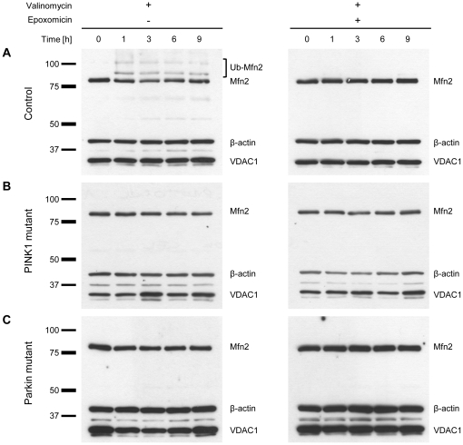
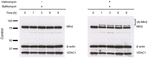
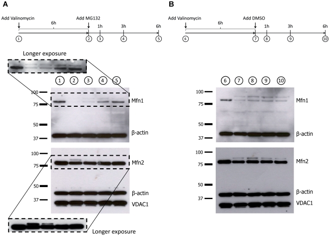
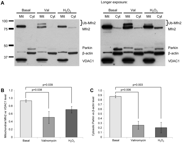
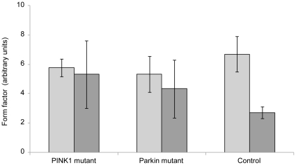
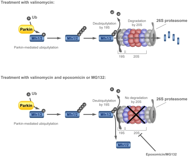
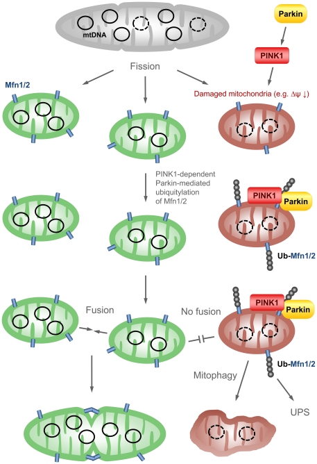
PINK1 and Parkin mutations cause recessive Parkinson's disease (PD). In Drosophila and SH-SY5Y cells, Parkin is recruited by PINK1 to damaged mitochondria, where it ubiquitinates Mitofusins and consequently promotes mitochondrial fission and mitophagy.Here, we investigated the impact of mutations in endogenous PINK1 and Parkin on the ubiquitination of mitochondrial fusion and fission factors and the mitochondrial network structure. Treating control fibroblasts with mitochondrial membrane potential (Δψ) inhibitors or H(2)O(2) resulted in ubiquitination of Mfn1/2 but not of OPA1 or Fis1. Ubiquitination of Mitofusins through the PINK1/Parkin pathway was observed within 1 h of treatment. Upon combined inhibition of Δψ and the ubiquitin proteasome system (UPS), no ubiquitination of Mitofusins was detected. Regarding morphological changes, we observed a trend towards increased mitochondrial branching in PD patient cells upon mitochondrial stress.For the first time in PD patient-derived cells, we demonstrate that mutations in PINK1 and Parkin impair ubiquitination of Mitofusins. In the presence of UPS inhibitors, ubiquitinated Mitofusin is deubiquitinated by the UPS but not degraded, suggesting that the UPS is involved in Mitofusin degradation.


Conflict of interest statement

Competing Interests: The authors have declared that no competing interests exist.

Figures

Figure 1
Figure 1. Expression of mitochondrial fusion and fission proteins after valinomycin treatment.
Fibroblasts from a healthy control, a homozygous PINK1 mutant and a homozygous Parkin mutant were cultured under basal conditions or treated with 1 µM valinomycin for 12 h. The protein levels of (A) Mfn2, (B) OPA1 and Fis1 were investigated by means of Western blotting. Valinomycin exposure caused a decrease in Mfn2 levels in controls, but not in PINK1- or Parkin-mutant cells. The protein levels of OPA1 and Fis1 were not influenced by this treatment in any of the cell cultures. Under basal and stress conditions, expression of the mitochondrial marker VDAC1 was comparable in mutants and controls. (C) Mutant cells were incubated with scrambled siRNA, Mfn1 siRNA, Mfn2 siRNA or a combination of Mfn1 and Mfn2 siRNA for 40 h. Western blot analysis was performed with an antibody against Mfn2. The Mfn2 level decreased only when Mfn2 siRNA was employed, confirming the specificity of the anti-Mfn1 antibody used in our study. β-actin served as a loading control. Fis1 – fission 1; Mfn1 – mitofusin 1; Mfn2 – mitofusin 2; OPA1 – optic atrophy 1; VDAC1 – voltage-dependent anion channel 1.
Figure 2
Figure 2. Ubiquitination of Mfn2 upon valinomycin treatment.
Fibroblasts from a healthy control were treated with 1 µM valinomycin for 6 h. Whole cell lysates from non-treated and valinomycin treated controls were immunoprecipitated using an antibody against Mfn2. Immunoprecipitates were analyzed by Western blotting using an antibody against ubiquitin (left panel). Subsequently, the membrane was washed and reprobed with an antibody against Mfn2 (right panel). Ubiquitinated forms of Mfn2 (mono- and polyubiquitinated) are present only in valinomycin treated controls. Mfn2 – mitofusin 2; Ub-Mfn2 – ubiquitinated mitofusin 2.
Figure 3
Figure 3. Mitochondrial localization of ubiquitinated Mfn2 after valinomycin treatment.
Control fibroblasts were cultured under basal conditions or treated with 1 µM valinomycin for 6 h. Cells were harvested and mitochondrial and cytosolic fractions were analyzed by Western blotting. The subcellular localization of Mfn2 was determined. Quality of cellular fractionation was confirmed using antibodies against VDAC1 and β-actin. The ubiquitinated forms of Mfn2, which were observed only after valinomycin stress, are exclusively found in the mitochondrial fraction. A longer exposure of the blot showed several Mfn2 bands with higher molecular weight, indicative of Mfn2 polyubiquitination (enlarged cutout). Cyt – cytosolic fraction; Mit – mitochondrial fraction; Mfn2 – mitofusin 2; VDAC1 – voltage-dependent anion channel 1.
Figure 4
Figure 4. Rescue of Mfn2 ubiquitination.
Fibroblasts of a control, a PINK1 and a Parkin mutant were transfected with an empty vector, a vector containing PINK1-V5 or a vector containing FLAG-Parkin. Twenty hours after transfection, cells were cultured under basal conditions or treated with 1 µM valinomycin for an additional 6 h. Western blot analysis using antibodies against V5 and FLAG showed bands of the size of full-length/cleaved PINK1 and Parkin, respectively, in treated and non-treated control cells after transfection, confirming the success of the experiment (upper panel). (A) In controls, ubiquitinated Mfn2 was detected in all three experiments after valinomycin treatment. (B) In PINK1-mutant cells, ubiquitination of Mfn2 under stress was rescued through overexpression of PINK1-V5 but not through overexpression of FLAG-Parkin. (C) In Parkin-mutant fibroblasts ubiquitinated Mfn2 was only detected after transfection with FLAG-Parkin. FL – full-length; Mfn2 – mitofusin 2; Ub-Mfn2 – ubiquitinated mitofusin 2.
Figure 5
Figure 5. Ubiquitination of Mfn2 is prevented by exposure to epoxomicin.
Fibroblasts from (A) a healthy control, (B) a homozygous PINK1 mutant and (C) a homozygous Parkin mutant were treated with 1 µM valinomycin alone (left panel) or with 1 µM valinomycin plus 10 µM epoxomicin (right panel). Proteins were extracted at different time points and analyzed by Western blotting. In control cells, valinomycin treatment initiated the ubiquitination of Mfn2 after 1 h of incubation. This effect was prevented by simultaneous exposure to epoxomicin. The mitochondrial marker VDAC1 and the cytosolic marker β-actin served as loading controls. Mfn2 – mitofusin 2; Ub-Mfn2 – ubiquitinated mitofusin 2; VDAC1 – voltage-dependent anion channel 1.
Figure 6
Figure 6. Exposure to bafilomycin has no impact on Mfn2 ubiquitination.
Control fibroblasts were treated with 10 nM bafilomycin alone (left panel) or with 10 nM bafilomycin plus 1 µM valinomycin (right panel). Proteins were extracted at different time points and analyzed by Western blotting. Bafilomycin had no effect on the Mfn2 levels and no ubiquitination was detected. When bafilomycin and valinomycin were combined, Mfn2 ubiquitination was initiated by valinomycin and not influenced by bafilomycin. In both experiments, the mitochondrial marker VDAC1 was unchanged. β-actin served as a loading control. Mfn2 – mitofusin 2; Ub-Mfn2 – ubiquitinated mitofusin 2; VDAC1 – voltage-dependent anion channel 1.
Figure 7
Figure 7. MG132 promotes deubiquitination of Mfn1 and Mfn2.
Control fibroblasts were treated with 1 µM valinomycin. After 6 h, MG132 (final concentration 10 µM) was added to the cells. Proteins were extracted at different time points and analyzed by Western blotting. Exposure to (A) an inhibitor of the UPS, i.e.MG132 but not to (B) DMSO alone, induced deubiquitination of both Mfn1 and Mfn2. Mfn1 – mitofusin 1; Mfn2 – mitofusin 2; VDAC1 – voltage-dependent anion channel 1.
Figure 8
Figure 8. Exposure to H2O2 causes ubiquitination of Mfn2.
Control fibroblasts were cultured under basal conditions, treated with 1 µM valinomycin for 6 h or with 100 µM H2O2 for 12 h. Cells were harvested and mitochondrial and cytosolic fractions were analyzed by Western blotting. (A) The subcellular localization of Mfn2 and Parkin was determined. Quality of cellular fractionation was confirmed using antibodies against VDAC1 and β-actin. (B) Densitometric analysis of the Western blot results revealed a significant drop in protein levels of non-modified Mfn2 (normalized against VDAC1 expression level) in the mitochondrial fraction after valinomycin or H2O2 treatment. (C) Furthermore, both treatments caused a significant drop in protein levels of Parkin (normalized against β-actin expression) in the cytosolic fraction. (A, right panel) A longer exposure of the Western blot shows mitochondrial translocation of Parkin after both treatments. For quantification of protein levels, blots of three independent experiments were evaluated. In the graphs mean intensities +/− standard deviation are given. Cyt – cytosolic fraction; H2O2 – hydrogen peroxide; Mit – mitochondrial fraction; Mfn2 – mitofusin 2; Ub-Mfn2 – ubiquitinated mitofusin 2; Val – Valinomycin; VDAC1 – voltage-dependent anion channel 1.
Figure 9
Figure 9. Branching of mitochondrial network.
The mitochondrial form factor was determined in a control, a homozygous PINK1 and a homozygous Parkin mutant. The quantification revealed no differences in mitochondrial branching between mutant and control cells under basal conditions (light gray bars). After treatment with 1 µM valinomycin for 12 h, a decrease in branching occurred in all individuals, with the control being most severely affected (dark gray bars). In the graph, mean form factors +/− standard deviation are given.
Figure 10
Figure 10. Possible scenario of Mfn1/2 deubiquitination and degradation at the UPS.
Under valinomycin stress, Mitofusins are ubiquitinated by Parkin. Poly-ubiquitinated Mitofusins are subsequently recognized by the intrinsic ubiquitin-binding activity of the 19S particle of the 26S proteasome. At the 19S regulatory complex the ubiquitin chain is disassembled, and the substrate is unfolded before it can enter the cavity of the 20S subunit where proteolysis takes place . Simultaneous treatment with epoxomicin (or MG132) inhibits the degradation function of the 20S core particle but does not influence the deubiquitylase activity of the 19S subunit. Consequently, poly-ubiquitinated Mitofusins are deubiquitinated at the proteasome but cannot be degraded. Mfn1/2 – mitofusin 1/2; Ub – ubiquitin; UPS – ubiquitin proteasome system.
Figure 11
Figure 11. Scheme summarizing PINK1's and Parkin's putative function in mitophagy.
By means of fission, mitochondria are randomly divided. Damaged mitochondria can be distinguished from functional mitochondria, for instance, by a difference in membrane potential. Dysfunctional mitochondria with low membrane potential are detected by PINK1, which recruits Parkin. Next, Parkin ubiquitinates Mfn1/2 which localize to the outer mitochondrial membrane. Subsequently, ubiquitinated Mitofusins are degraded by the UPS, preventing fusion of dysfunctional with functional mitochondria. By this, dysfunctional mitochondria are selected out from the general pool of mitochondria and subsequently undergo mitophagy. Δψ – mitochondrial membrane potential; Mfn1/2 – mitofusin 1/2; PINK1 – PTEN-induced putative kinase 1; Ub-Mfn1/2 – ubiquitinated mitofusin 1/2; UPS – ubiquitin proteasome system.

References

    1. Klein C, Schlossmacher MG. Parkinson disease, 10 years after its genetic revolution: multiple clues to a complex disorder. Neurology. 2007;69:2093–2104. - PubMed
    1. Palacino JJ, Sagi D, Goldberg MS, Krauss S, Motz C, et al. Mitochondrial dysfunction and oxidative damage in parkin-deficient mice. J Biol Chem. 2004;279:18614–18622. - PubMed
    1. Flinn L, Mortiboys H, Volkmann K, Koster RW, Ingham PW, et al. Complex I deficiency and dopaminergic neuronal cell loss in parkin-deficient zebrafish (Danio rerio). Brain. 2009;132:1613–1623. - PubMed
    1. Gegg ME, Cooper JM, Schapira AH, Taanman JW. Silencing of PINK1 expression affects mitochondrial DNA and oxidative phosphorylation in dopaminergic cells. PLoS One. 2009;4:e4756. - PMC - PubMed
    1. Schapira AH, Cooper JM, Dexter D, Jenner P, Clark JB, et al. Mitochondrial complex I deficiency in Parkinson's disease. Lancet. 1989;1:1269. - PubMed

Publication types

MeSH terms

Substances