. 2011 Mar 8;6(2):e17344.
doi: 10.1371/journal.pone.0017344.

New modularity of DAP-kinases: alternative splicing of the DRP-1 gene produces a ZIPk-like isoform

Affiliations

New modularity of DAP-kinases: alternative splicing of the DRP-1 gene produces a ZIPk-like isoform

Yishay Shoval et al. PLoS One. .

Abstract

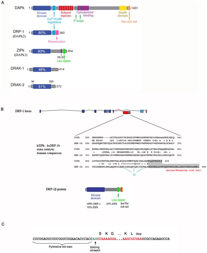
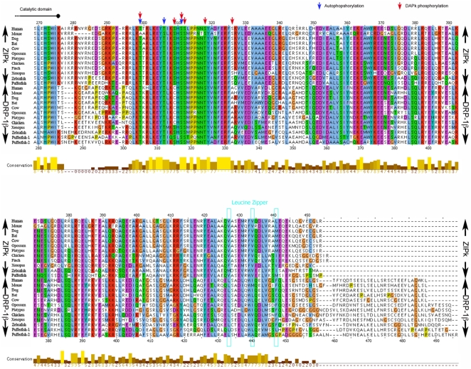
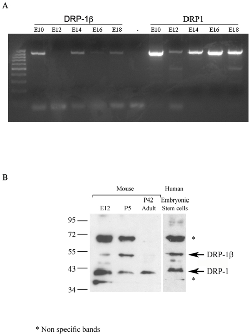
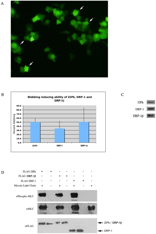
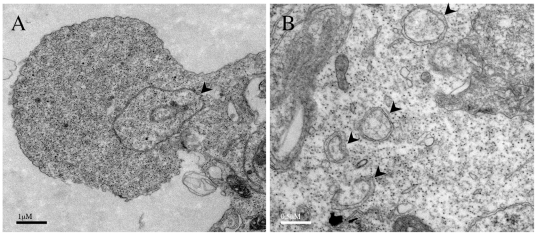
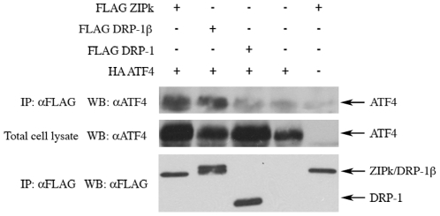
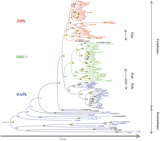
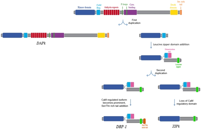
DRP-1 and ZIPk are two members of the Death Associated Protein Ser/Thr Kinase (DAP-kinase) family, which function in different settings of cell death including autophagy. DAP kinases are very similar in their catalytic domains but differ substantially in their extra-catalytic domains. This difference is crucial for the significantly different modes of regulation and function among DAP kinases. Here we report the identification of a novel alternatively spliced kinase isoform of the DRP-1 gene, termed DRP-1β. The alternative splicing event replaces the whole extra catalytic domain of DRP-1 with a single coding exon that is closely related to the sequence of the extra catalytic domain of ZIPk. As a consequence, DRP-1β lacks the calmodulin regulatory domain of DRP-1, and instead contains a leucine zipper-like motif similar to the protein binding region of ZIPk. Several functional assays proved that this new isoform retained the biochemical and cellular properties that are common to DRP-1 and ZIPk, including myosin light chain phosphorylation, and activation of membrane blebbing and autophagy. In addition, DRP-1β also acquired binding to the ATF4 transcription factor, a feature characteristic of ZIPk but not DRP-1. Thus, a splicing event of the DRP-1 produces a ZIPk like isoform. DRP-1β is highly conserved in evolution, present in all known vertebrate DRP-1 loci. We detected the corresponding mRNA and protein in embryonic mouse brains and in human embryonic stem cells thus confirming the in vivo utilization of this isoform. The discovery of module conservation within the DAPk family members illustrates a parsimonious way to increase the functional complexity within protein families. It also provides crucial data for modeling the expansion and evolution of DAP kinase proteins within vertebrates, suggesting that DRP-1 and ZIPk most likely evolved from their ancient ancestor gene DAPk by two gene duplication events that occurred close to the emergence of vertebrates.


Conflict of interest statement

Competing Interests: The authors have declared that no competing interests exist.

Figures

Figure 1
Figure 1. The DAPk family of proteins and the new member, DRP-1β.
A. The percentage in the blue boxes, representing the catalytic domain of the kinases, indicates the extent of identity of each catalytic domain to the kinase domain of DAPk. B. A scheme of the genomic locus of DRP-1, DRP-1β exon and the DRP-1β protein structure. Dark blue- catalytic domain coding exons; light blue- CaM binding domain encoding exons, pink- dimerization tail encoding exons; red and green- the alternative open reading frame. Percents indicate similarity of the catalytic or extra-catalytic domain to the indicated protein. Enlarged area shows sequence alignment of the human alternative exon and the extra catalytic domain of human ZIPk. Letters indicate identities, pluses indicate similarities. Gray background indicates a non aligned area. LZ- leucine zipper. C. DNA sequence at the 5′ and 3′ of human DRP-1β alternative exon. Red- open reading frame; Green- splicing acceptor site. Capital letters- translated amino acids.
Figure 2
Figure 2. The DRP-1β alternative exon shows similarity to the extra-catalytic domain of ZIPk.
A multiple sequence alignments of the extra catalytic domain of ZIPk and DRP-1β orthologs from the indicated vertebrates. Blue arrows- ZIPk autophosphorylation sites; red arrows- ZIPk phosphorylation sites by DAPk. Brown to yellow bars- conservation measure; the position of ZIPk leucine zipper is marked by pale blue boxes. The MSA was performed using CLUSTALW program and visualized with the JalView tool.
Figure 3
Figure 3. mRNA and protein expression of DRP-1β.
A. DRP-1β and DRP-1 mRNA fragments were amplified by PCR, using total embryo mouse cDNA from the indicated days as template, followed by ethidium bromide gel detection. B. Western blot detection of DRP-1β and DRP-1 protein levels in brain tissues of mice and human embryonic stem cells, using anti N'-DRP-1 antibody. E12- embryonic day 12, P5 - postnatal day 5, P42 – postnatal day 42 (adult mouse).
Figure 4
Figure 4. Ectopically expressed DRP-1β induces MLC phosphorylation and membrane blebbing in cells.
A. DRP-1β ectopic expression induces membrane blebbing. HEK293T cells were co-transfected with FLAG DRP-1β and GFP expression vectors, and examined under fluorescent microscope after 24 h. White arrows- cells exhibiting membrane blebs. B. Quantification of the blebbing inducing ability of ZIPk, DRP-1 and DRP-1β. Note that the number of blebbed cells in cells transfested with control plasmids is below detection levels C. Western blot detection of the kinases, (detection was done with anti-FLAG Abs, running the samples in the same gels and the same exposure time of the blots) indicating comparable expression levels. D. ZIPk, DRP-1β and DRP-1 phosphorylate myosin light chain (MLC). FLAG tagged kinases were expressed in HEK293T, immunoprecipitated using anti-FLAG antibodies and eluted from beads. His-tagged MLC was purified from bacteria, and used as a substrate in an in vitro kinase assay. MLC phosphorylation was detected using an antibody against phospho-serine 19 on MLC.
Figure 5
Figure 5. Ectopic expression of DRP-1β induces the accumulation of autophagic vesicles.
HEK293T cells were transfected with DRP-1β expression vector, fixed 24 h after transfection and examined using Transmission Electron Microscopy (TEM). A. a cell undergoing membrane blebbing; B. larger magnification of induced vesicles. Arrowheads indicate double membrane, autophagic vesicles.
Figure 6
Figure 6. ZIPk and DRP-1β bind ATF-4, while DRP-1 fails to do so.
HEK293T cells were co-transfected with the indicated vectors and harvested 24 h post transfection. Lysates were immunoprecipitated using anti-FLAG antibodies, and protein levels were detected using western blot.
Figure 7
Figure 7. The DAP kinases phylogenetic tree.
A phylogenetic tree of the indicated organisms was constructed based on the multiple alignment of the DAP kinase scatalytic domain, using the PHYML program. Numbers above branches represent bootstrap support from 100 replicates. Yellow background- high bootstraps value. Blue- DAPk; Green- DRP-1; Red- ZIPk; Black- ortholog undetermined due to partial sequence.
Figure 8
Figure 8. A model of the Evolution of the DAP kinases.
Scheme showing a most-parsimonious model of the evolution of DRP-1 and ZIPk in vertebrates.

References

    1. Bialik S, Kimchi A. The death-associated protein kinases: structure, function, and beyond. Annu Rev Biochem. 2006;75:189–210. - PubMed
    1. Shohat G, Shani G, Eisenstein M, Kimchi A. The DAP-kinase family of proteins: study of a novel group of calcium-regulated death-promoting kinases. Biochim Biophys Acta. 2002;1600:45–50. - PubMed
    1. Tu W, Xu X, Peng L, Zhong X, Zhang W, et al. DAPK1 interaction with NMDA receptor NR2B subunits mediates brain damage in stroke. Cell. 2010;140:222–234. - PMC - PubMed
    1. Bialik S, Kimchi A. DAP-kinase as a target for drug design in cancer and diseases associated with accelerated cell death. Semin Cancer Biol. 2004;14:283–294. - PubMed
    1. Esteller M. Relevance of DNA methylation in the management of cancer. Lancet Oncol. 2003;4:351–358. - PubMed

Publication types

MeSH terms

Substances