. 2011 Mar 7;6(3):e17487.
doi: 10.1371/journal.pone.0017487.

Accumulation of DNA damage in hematopoietic stem and progenitor cells during human aging

Affiliations

Accumulation of DNA damage in hematopoietic stem and progenitor cells during human aging

Claudia E Rübe et al. PLoS One. .

Abstract

Background: Accumulation of DNA damage leading to adult stem cell exhaustion has been proposed to be a principal mechanism of aging. Here we tested this hypothesis in healthy individuals of different ages by examining unrepaired DNA double-strand breaks (DSBs) in hematopoietic stem/progenitor cells matured in their physiological microenvironment.

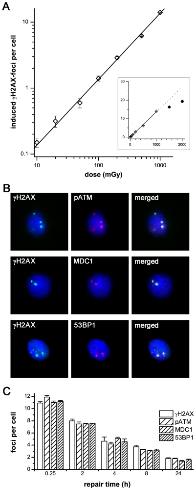
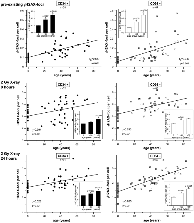
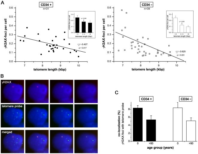
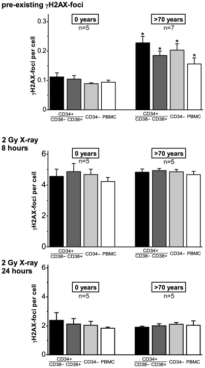
Methodology/principal findings: To asses DNA damage accumulation and repair capacities, γH2AX-foci were examined before and after exposure to ionizing irradiation. Analyzing CD34+ and CD34- stem/progenitor cells we observed an increase of endogenous γH2AX-foci levels with advancing donor age, associated with an age-related decline in telomere length. Using combined immunofluorescence and telomere-fluorescence in-situ hybridization we show that γH2AX-foci co-localize consistently with other repair factors such as pATM, MDC1 and 53BP1, but not significantly with telomeres, strongly supporting the telomere-independent origin for the majority of foci. The highest inter-individual variations for non-telomeric DNA damage were observed in middle-aged donors, whereas the individual DSB repair capacity appears to determine the extent of DNA damage accrual. However, analyzing different stem/progenitor subpopulations obtained from healthy elderly (>70 years), we observed an only modest increase in DNA damage accrual, most pronounced in the primitive CD34+CD38(-)-enriched subfraction, but sustained DNA repair efficiencies, suggesting that healthy lifestyle may slow down the natural aging process.

Conclusions/significance: Based on these findings we conclude that age-related non-telomeric DNA damage accrual accompanies physiological stem cell aging in humans. Moreover, aging may alter the functional capacity of human stem cells to repair DSBs, thereby deteriorating an important genome protection mechanism leading to exceeding DNA damage accumulation. However, the great inter-individual variations in middle-aged individuals suggest that additional cell-intrinsic mechanisms and/or extrinsic factors contribute to the age-associated DNA damage accumulation.


Conflict of interest statement

Competing Interests: The authors have declared that no competing interests exist.

Figures

Figure 1
Figure 1. γH2AX-foci in hematopoietic stem and progenitor cells.
A: γH2AX-foci in CD34+ cells were enumerated at 0.5 h after irradiation with doses from 10 mGy to 2000 mGy and plotted against the irradiation dose. The induction of γH2AX-foci is clearly dependent on the irradiation dose, with a linear correlation from 10 mGy to 1000 mGy. At foci levels of more than 15 foci per cell, the clustering of foci impedes their clear discrimination, leading to an underestimation of actual foci numbers (inset). B: Immunofluorescence double-staining of γH2AX (green) combined with pATM, MDC1 or 53BP1 in CD34+ cells, 8 h after irradiation with 2 Gy. The clear co-localization with other DNA repair factors confirms that γH2AX-foci can be used to analyze DSBs. (Original magnification, ×600) C: Quantitative analysis of γH2AX-, pATM-, MDC1- and 53BP1-foci in CD34+ cells at different time-points after irradiation with 1 Gy. γH2AX-foci co-localize consistently with other DNA repair factors. Error bars signify the SE of three different experiments.
Figure 2
Figure 2. Accumulation of DSBs and declining DSB repair capacities with advancing age.
The number of pre-existing γH2AX-foci (upper panels), and radiation-induced γH2AX-foci at 8 hours (middle panel) and 24 hours (lower panels) after irradiation with 2 Gy was plotted against donor age, depicted separately for CD34+ and CD34− cells. Linear regression analyses were performed (solid lines) and Spearman's rank correlation coefficients (rs) were calculated. For three different arbitrary groups (age: 0/<50/>50 years) mean γH2AX-foci levels are displayed in bar graphs. Error bars represent the SE within the specific group. *Statistically significant difference to the next younger age group (p<0.05).
Figure 3
Figure 3. Accumulation of DSBs and progressive telomere-shortening.
A: The number of pre-existing γH2AX-foci per cell was plotted against telomere length depicted separately for CD34+ and CD34− cells. Linear regression analyses were performed (solid lines) and Spearman's rank correlation coefficients (rs) were calculated. For three different arbitrary groups (telomere length: <8/8–9/>9 kb) mean γH2AX-foci levels are displayed in bar graphs. Error bars represent the SE within the specific group. *Statistically significant difference to the next younger age group or next group with shorter telomere (p<0.05). B: Co-localization between γH2AX- and telomere-signals. Co-localization of endogenous γH2AX-foci (red) with telomere-specific DNA (green FISH signal). DAPI staining (blue) indicates DNA. (Original magnification, ×600) C: Quantitative analysis of the extent of co-localization between γH2AX- and telomere-signals. In CD34+ and CD34− cells obtained from cord blood and bone marrow of aged individuals (>50 years), respectively, approximately 200 γH2AX-foci were screened per data point in independent experiments. Error bars signify the SE of five different experiments.
Figure 4
Figure 4. Accumulation of DSBs in the different stem/progenitor populations during healthy aging.
The number of pre-existing γH2AX-foci (upper panel), and radiation-induced γH2AX-foci at 8 hours (middle panel) and 24 hours (lower panel) after irradiation with 2 Gy was counted in CD34+CD38−, CD34+CD38+, CD34− and PBMC cells derived from cord blood (0 years) and bone marrow of healthy elderly (>70 years). Error bars represent the SE within the specific group. *Statistically significant difference to the corresponding cord-blood derived stem/progenitor population (p<0.05).

References

    1. Hoeijmakers JH. DNA damage, aging, and cancer. N Engl J Med. 2009;361:1475–1485. - PubMed
    1. Rogakou EP, Pilch DR, Orr AH, Ivanova VS, Bonner WM. DNA double-stranded breaks induce histone H2AX phosphorylation on serine 139. J Biol Chem. 1998;273:5858–5868. - PubMed
    1. Rothkamm K, Lobrich M. Evidence for a lack of DNA double-strand break repair in human cells exposed to very low x-ray doses. Proc Natl Acad Sci U S A. 2003;100:5057–5062. - PMC - PubMed
    1. Rube CE, Dong X, Kuhne M, Fricke A, Kaestner L, et al. DNA double-strand break rejoining in complex normal tissues. Int J Radiat Oncol Biol Phys. 2008;72:1180–1187. - PubMed
    1. Rube CE, Grudzenski S, Kuhne M, Dong X, Rief N, et al. DNA double-strand break repair of blood lymphocytes and normal tissues analysed in a preclinical mouse model: implications for radiosensitivity testing. Clin Cancer Res. 2008;14:6546–6555. - PubMed

Publication types