. 2011 Mar;7(3):e1002010.
doi: 10.1371/journal.pgen.1002010. Epub 2011 Mar 3.

A bow-tie genetic architecture for morphogenesis suggested by a genome-wide RNAi screen in Caenorhabditis elegans

Affiliations

A bow-tie genetic architecture for morphogenesis suggested by a genome-wide RNAi screen in Caenorhabditis elegans

Matthew D Nelson et al. PLoS Genet. 2011 Mar.

Abstract

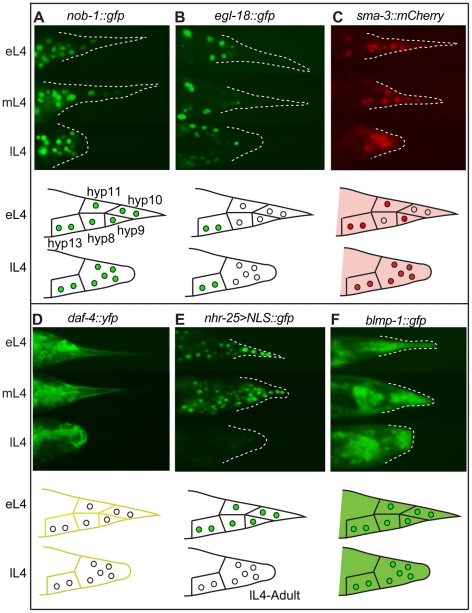
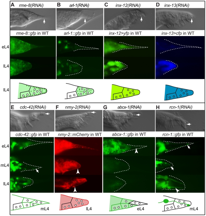
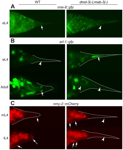
During animal development, cellular morphogenesis plays a fundamental role in determining the shape and function of tissues and organs. Identifying the components that regulate and drive morphogenesis is thus a major goal of developmental biology. The four-celled tip of the Caenorhabditis elegans male tail is a simple but powerful model for studying the mechanism of morphogenesis and its spatiotemporal regulation. Here, through a genome-wide post-embryonic RNAi-feeding screen, we identified 212 components that regulate or participate in male tail tip morphogenesis. We constructed a working hypothesis for a gene regulatory network of tail tip morphogenesis. We found regulatory roles for the posterior Hox genes nob-1 and php-3, the TGF-β pathway, nuclear hormone receptors (e.g. nhr-25), the heterochronic gene blmp-1, and the GATA transcription factors egl-18 and elt-6. The majority of the pathways converge at dmd-3 and mab-3. In addition, nhr-25 and dmd-3/mab-3 regulate each others' expression, thus placing these three genes at the center of a complex regulatory network. We also show that dmd-3 and mab-3 negatively regulate other signaling pathways and affect downstream cellular processes such as vesicular trafficking (e.g. arl-1, rme-8) and rearrangement of the cytoskeleton (e.g. cdc-42, nmy-1, and nmy-2). Based on these data, we suggest that male tail tip morphogenesis is governed by a gene regulatory network with a bow-tie architecture.


Conflict of interest statement

The authors have declared that no competing interests exist.

Figures

Figure 1
Figure 1. Overview of tail tip morphogenesis and the experimental design for the genome-wide RNAi screen for tail tip defects in male C. elegans.
(A) Schematic of tail tip morphogenesis: the tail tip cells hyp8–hyp11 fuse and then detach from the overlying cuticle. The newly formed syncytium changes shape—becoming rounded—and migrates anteriorly. By the adult stage, the male tail has flattened out ventrally. (B) Screening methodology: L1 larvae were fed dsRNA-producing bacteria on thin films of nutrient agar and grown to adulthood. A square of agar containing worms was mounted on a slide and scored at 400× magnification. (C) The phenotypes scored included WT (wild type), Lep (leptoderan), and Ore (over-retracted). Additional phenotypes scored but not reported here were spicule morphology and general male tail defects (see http://wormtails.bio.nyu.edu). (D) Distribution of the phenotypes among the 212 positive genes; for 8 genes, Lep and Ore tails were observed in the same experiment. GO attribute enrichment via FuncAssociate for genes causing Lep, Ore and Lep+Ore phenotypes upon RNAi. For each phenotype, the top six categories that were over-represented are indicated with bar graphs depicting the LOD scores, i.e. the log of the odds that the frequency of a GO category is no different than that for all genes in the genome.
Figure 2
Figure 2. Expression patterns of sample regulatory genes identified in the RNAi screen.
Fluorescent micrographs for early, middle and late L4 larvae and schematic drawings for early and late L4 larvae are shown in the upper and lower portions of each panel, respectively. (A) NOB-1::GFP is expressed in hyp8–11 throughout larval development. (B) EGL-18::GFP is expressed in the male tail but not in hyp8–11. (C) SMA-3::mCherry is cytoplasmic in early L4 and begins to accumulate in the nuclei of the tail tip cells. It then remains nuclear and cytoplasmic throughout tail tip morphogenesis. (D) DAF-4::YFP localizes to the membranes of hyp8–11 prior to and during morphogenesis. (E) An nhr-25 transcriptional reporter shows expression in hyp8–11 at the beginning of morphogenesis, followed by a rapid shutoff. (F) BLMP-1::GFP is expressed in the cytoplasm and nuclei of hyp8–11 throughout larval development.
Figure 3
Figure 3. Genes with Ore-like phenotypes.
(A) DIC images of male tails: RNAi of smc-4 resulted in Ore (arrow) and Lep (arrowhead) phenotypes. npp-3 RNAi resulted in severe Ore tails (arrow). (B) Subnetwork of genes that produce the Ore phenotype as reconstructed by N-Browse . Thirteen of the 22 genes with an Ore phenotype are tightly clustered: purple, Ore RNAi phenotype; green, Lep RNAi phenotype; black edges: predicted genetic interactions; orange edges: expression correlation; blue edges: phenotypic correlation in the embryo; green edges: protein-protein interaction (multiple data sources, see [17]).
Figure 4
Figure 4. Effectors of morphogenesis identified in the RNAi screen.
DIC images of RNAi phenotypes in adult males (top of each panel), expression patterns in the male tail of early L4 (eL4), mid L4 (mL4) and/or late L4 (lL4) (middle of each panel), and schematic of expression/localization patterns (bottom of each panel). Arrows in the DIC images indicate Lep tail tips; arrows in the fluorescent images indicate localization patterns of interest. (A) rme-8(RNAi) results in a total failure of tail tip morphogenesis. RME-8::GFP localizes to the edge of detaching and retracting tail tip cells as puncta (depicted as dark green dots in the schematic). (B) arl-1(RNAi) causes a disruption of tail tip retraction. ARL-1::GFP is expressed cytoplasmically at extremely low levels in hyp8–11 prior to morphogenesis (eL4); during morphogenesis (lL4), it localizes to distinct puncta (dark green dots in the schematic). (C, D) inx-12(RNAi) and inx-13(RNAi) result in subtle Lep phenotypes. Reporters for inx-12 and inx-13 are expressed in tail tip cells prior to and during morphogenesis. (E) cdc-42(RNAi) results in a Lep phenotype. CDC-42::GFP is cytoplasmic prior to tail tip retraction, localizes to the membranes and discrete puncta during retraction (arrow), and becomes cytoplasmic again in late L4 and adults. (F) nmy-2(RNAi) results in Lep phenotypes. NMY-2::mCherry localizes to the posterior end of retracting tail tip cells and to the ventral surface of the male tail (arrowheads). (G) abcx-1(RNAi) results in Lep phenotypes. ABCX-1::GFP is expressed in hyp8, 9 and 11 prior to but not during morphogenesis. Expression is absent from hyp10 (arrowhead). (H) rcn-1(RNAi) causes Lep phenotypes. RCN-1::GFP is expressed in PHCL and PHCR (arrows), as well as in other more anterior neurons during tail tip morphogenesis. After morphogenesis, expression is intense in the phasmid socket cells (arrowhead).
Figure 5
Figure 5. A partial genetic network of tail tip morphogenesis as predicted by N-Browse (thin lines) and other published work (thick dashed lines) , , –, .
Clusters of pathways or functional groups are surrounded by boxes to represent their proximity within the network and/or functional interactions in modules. Nodes (genes): green, Lep positives identified in the screen; purple, Ore positives identified in the screen; green/purple, Lep+Ore positives from the screen; yellow, previously identified tail tip genes; green boundary (unfilled), not identified in the screen but suggested by the network and experimentally validated; red boundary (unfilled), not positive in the screen and not further tested. Edges (lines) represent the following types of data concerning interactions: dashed black: published and experimentally validated interactions; blue: interologs (conserved interactions between pairs of proteins that have interacting homologs in other organisms); gray: predicted genetic interactions; purple: experimentally verified genetic interactions; green: protein-protein interactions; orange: expression correlations.
Figure 6
Figure 6. Expression epistasis experiments to test interactions between key regulatory genes.
In the top of each panel are fluorescent micrographs of male tails (lateral views) at three of several stages (eL3 =  early L3, lL3 =  late L3, eL4 =  early L4, mL4 =  mid-L4, lL4 =  late L4, or adult) expressing various transgenes in different genetic backgrounds. At the bottom of each panel is a schematic depicting the expression pattern at one exemplar stage. (A) Expression of a dmd-3>yfp transcriptional reporter in wild-type males. (B–G) Effects on the expression of dmd-3>yfp by RNAi-depletion or mutations of other genes (note that tail tip morphogenesis is impaired in these RNAi-treated or mutant males and rounding of the tail tip in lL4 does not occur). (B) Loss of php-3 function in ok919 mutant animals causes reduced dmd-3>yfp expression in hyp8, hyp11 and hyp13. (C) Depletion of nob-1 by RNAi causes reduced dmd-3>yfp expression in hyp8–11. (D) php-3(ok919) nob-1(RNAi) animals show no dmd-3>yfp expression in hyp8-11. (E) Although dmd-3>yfp is initially expressed in egl-18(ok290) elt-6(RNAi) animals, it shuts off prematurely in hyp8–11. (F) Expression of dmd-3>yfp is never initiated in the tail tip cells of nhr-25(ku217) reduced-function mutants. (G) blmp-1(tm548) mutants show precocious expression of dmd-3>yfp in eL3 in hyp8, hyp9 and hyp11, no expression in hyp10, and premature inactivation of dmd-3>yfp at early L4. (H) nhr-25>NLS::gfp is expressed brightly prior to and at the beginning of morphogenesis and is quickly inactivated near its end. (I) In mab-3(e1240);dmd-3(ok1327) animals, expression of the nhr-25 reporter remains bright in adult males. (J) SMA-3::CFP is expressed and nuclearly localized in hyp8–11 during morphogenesis and is not visible in adult males (the image shows autofluorescence of the spicules and the fan). (K) In mab-3(e1240);dmd-3(ok1327) adults, expression of the sma-3::cfp reporter remains very bright into adulthood. (L) vav-1>NLS::gfp is expressed brightly in hyp8–11 and in other cells of the tail prior to and during morphogenesis and at extremely low levels in adults. (M) Expression of the vav>NLS::gfp reporter remains very bright in hyp8–11 and the other cells of the male tail in adult mab-3(e1240);dmd-3(ok1327) animals.
Figure 7
Figure 7. Cellular processes controlled by DMD-3 and MAB-3.
Expression patterns of translational fusions are shown for wild-type males (left side of each panel) and mab-3(e1240);dmd-3(ok1327) mutant males (right side of each panel). (A) DMD-3 and MAB-3 regulate RME-8 localization but not expression. In wild-type animals, RME-8::GFP localizes as discrete puncta to the cell cortex where the tail tip cells are detaching from the cuticle (arrow), but is dispersed and cytoplasmic (arrowhead) in mab-3(e1240);dmd-3(ok1327) mutants. (B) In early L4, ARL-1::GFP shows very low levels of expression in wild-type males (arrowhead) but bright expression in hyp10 (arrow) in mab-3(e1240);dmd-3(ok1327) males. In adults, ARL-1::GFP is expressed in tail neurons in wild-type males (arrow points to phasmids), but not in mab-3(e1240);dmd-3(ok1327) males (arrowheads). (C) In wild-type males, NMY-2::mCherry localization is focused to a cap at the posterior end of the tail tip during rounding and to the region along the ventral edge where the tail tip cells are detaching from the cuticle (arrows). In mab-3(e1240);dmd-3(ok1327) animals, localization to the ventral surface still occurs (arrows), but localization to the posterior end of the tail is not observed (arrowheads).
Figure 8
Figure 8. The genetic architecture of tail tip morphogenesis, showing experimentally validated connections between components.
This network has a bow-tie architecture with dmd-3, mab-3 and nhr-25 in the core. The central role for dmd-3 and mab-3 was originally proposed by Mason et al. . We found that nhr-25 is required for the activation of dmd-3 and forms a negative feedback loop with dmd-3 and mab-3. Multiple signaling pathways/genes feed into the core by regulating dmd-3 positively (lines with arrow at one end) or negatively (lines with bar at one end). Genes controlling morphogenesis (rme-8, nmy-2, arl-1, eff-1) and other signaling pathways (TGF-β and vav-1-mediated) are regulated by dmd-3 and mab-3. Dashed lines indicate possibly direct or indirect interactions, solid lines indicate an experimentally validated direct interactions .

Similar articles

Cited by

References

    1. Hadjantonakis K, Solnica-Krezel L. Developmental biology 50 years-investigating the emergence of shape. Introduction. Dev Biol. 2010;341:2–4. - PMC - PubMed
    1. Sulston JE, Albertson DG, Thomson JN. The Caenorhabditis elegans male: postembryonic development of nongonadal structures. Dev Biol. 1980;78:542–576. - PubMed
    1. Nguyen CQ, Hall DH, Yang Y, Fitch DH. Morphogenesis of the Caenorhabditis elegans male tail tip. Dev Biol. 1999;207:86–106. - PubMed
    1. Sulston JE, Horvitz HR. Post-embryonic cell lineages of the nematode, Caenorhabditis elegans. Dev Biol. 1977;56:110–156. - PubMed
    1. Zhao X, Yang Y, Fitch DH, Herman MA. TLP-1 is an asymmetric cell fate determinant that responds to Wnt signals and controls male tail tip morphogenesis in C. elegans. Development. 2002;129:1497–1508. - PubMed

Publication types

MeSH terms

Substances

LinkOut - more resources