

Evaluation of phototoxicity of dendritic porphyrin-based phosphorescent oxygen probes: an in vitro study
- PMID: 21409208
- PMCID: PMC3607943
- DOI: 10.1039/c0pp00356e
Evaluation of phototoxicity of dendritic porphyrin-based phosphorescent oxygen probes: an in vitro study
Abstract
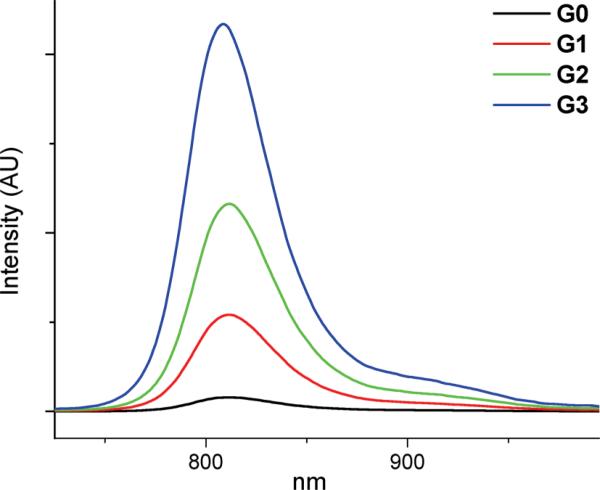
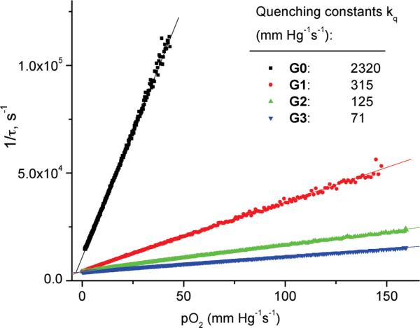
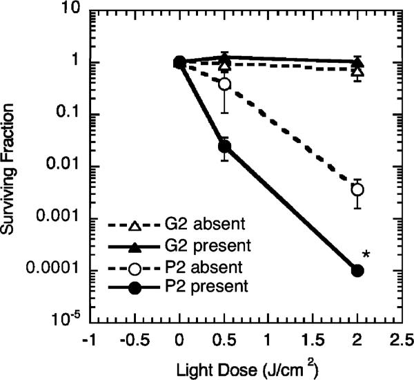
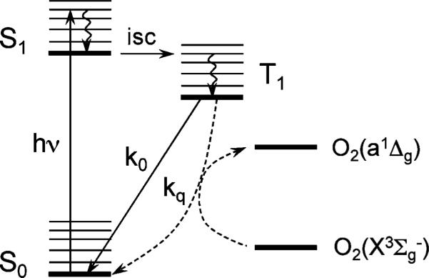
Biological oxygen measurements by phosphorescence quenching make use of exogenous phosphorescent probes, which are introduced directly into the medium of interest (e.g. blood or interstitial fluid) where they serve as molecular sensors for oxygen. The byproduct of the quenching reaction is singlet oxygen, a highly reactive species capable of damaging biological tissue. Consequently, potential probe phototoxicity is a concern for biological applications. Herein, we compared the ability of polyethyleneglycol (PEG)-coated Pd tetrabenzoporphyrin (PdTBP)-based dendritic nanoprobes of three successive generations to sensitize singlet oxygen. It was found that the size of the dendrimer has practically no effect on the singlet oxygen sensitization efficiency in spite of the strong attenuation of the triplet quenching rate with an increase in the dendrimer generation. This unexpected result is due to the fact that the lifetime of the PdTBP triplet state in the absence of oxygen increases with dendritic generation, thus compensating for the concomitant decrease in the rate of quenching. Nevertheless, in spite of their ability to sensitize singlet oxygen, the phosphorescent probes were found to be non-phototoxic when compared with the commonly used photodynamic drug Photofrin in a standard cell-survival assay. The lack of phototoxicity is presumably due to the inability of PEGylated probes to associate with cell surfaces and/or penetrate cellular membranes. In contrast, conventional photosensitizers bind to cell components and act by generating singlet oxygen inside or in the immediate vicinity of cellular organelles. Therefore, PEGylated dendritic probes are safe to use for tissue oxygen measurements as long as the light doses are less than or equal to those commonly employed in photodynamic therapy.
Figures


Similar articles
-
The phototoxicity of photofrin was enhanced by PEGylated liposome in vitro.Cancer Lett. 2006 Sep 8;241(1):42-8. doi: 10.1016/j.canlet.2005.10.024. Epub 2005 Nov 21. Cancer Lett. 2006. PMID: 16303246
-
Dendritic phosphorescent probes for oxygen imaging in biological systems.ACS Appl Mater Interfaces. 2009 Jun;1(6):1292-304. doi: 10.1021/am9001698. ACS Appl Mater Interfaces. 2009. PMID: 20072726 Free PMC article.
-
Efficient intersystem crossing using singly halogenated carbomethoxyphenyl porphyrins measured using delayed fluorescence, chemical quenching, and singlet oxygen emission.Phys Chem Chem Phys. 2015 Nov 21;17(43):29090-6. doi: 10.1039/c5cp04359j. Phys Chem Chem Phys. 2015. PMID: 26460933
-
Accessibility and external versus intercalative binding to DNA as assessed by oxygen-induced quenching of the palladium(II)-containing cationic porphyrins Pd(T4) and Pd(tD4).Biochemistry. 2014 Feb 4;53(4):714-24. doi: 10.1021/bi401610t. Epub 2014 Jan 23. Biochemistry. 2014. PMID: 24428500
-
Porphyrin-Containing MOFs and COFs as Heterogeneous Photosensitizers for Singlet Oxygen-Based Antimicrobial Nanodevices.ACS Appl Mater Interfaces. 2021 Jun 16;13(23):26651-26672. doi: 10.1021/acsami.1c05234. Epub 2021 Jun 4. ACS Appl Mater Interfaces. 2021. PMID: 34086450 Review.
Cited by
-
Ultrafast Tracking of Oxygen Dynamics During Proton FLASH.Int J Radiat Oncol Biol Phys. 2022 Jul 1;113(3):624-634. doi: 10.1016/j.ijrobp.2022.03.016. Epub 2022 Mar 18. Int J Radiat Oncol Biol Phys. 2022. PMID: 35314293 Free PMC article.
-
Dual-Emissive Difluoroboron Naphthyl-Phenyl β-Diketonate Polylactide Materials: Effects of Heavy Atom Placement and Polymer Molecular Weight.Macromolecules. 2014 Jun 10;47(11):3736-3746. doi: 10.1021/ma5006606. Epub 2014 May 23. Macromolecules. 2014. PMID: 24954954 Free PMC article.
-
Optical probes and techniques for O2 measurement in live cells and tissue.Cell Mol Life Sci. 2012 Jun;69(12):2025-39. doi: 10.1007/s00018-011-0914-0. Epub 2012 Jan 17. Cell Mol Life Sci. 2012. PMID: 22249195 Free PMC article. Review.
-
Direct Measurements of FLASH-Induced Changes in Intracellular Oxygenation.Int J Radiat Oncol Biol Phys. 2024 Mar 1;118(3):781-789. doi: 10.1016/j.ijrobp.2023.09.019. Epub 2023 Sep 18. Int J Radiat Oncol Biol Phys. 2024. PMID: 37729972 Free PMC article.
-
Oxyphor 2P: A High-Performance Probe for Deep-Tissue Longitudinal Oxygen Imaging.Cell Metab. 2019 Mar 5;29(3):736-744.e7. doi: 10.1016/j.cmet.2018.12.022. Epub 2019 Jan 24. Cell Metab. 2019. PMID: 30686745 Free PMC article.
References
-
- Vanderkooi JM, Maniara G, Green TJ, Wilson DF. An optical method for measurement of dioxygen concentration based on quenching of phosphorescence. J. Biol. Chem. 1987;262:5476–5482. - PubMed
-
- Wilson DF. Quantifying the role of oxygen pressure in tissue function. Am. J. Physiol.: Heart Circ. Physiol. 2007;294:H11–H13. - PubMed
-
- Mik EG, Johannes T, Ince C. Monitoring of renal venous Po-2 and kidney oxygen consumption in rats by a near-infrared phosphorescence lifetime technique. Am. J. Physiol.: Renal Physiol. 2008;294:F676–F681. - PubMed
-
- Golub AS, Barker MC, Pittman RN. Microvascular oxygen tension in the rat mesentery. Am. J. Physiol.: Heart Circ. Physiol. 2007;294:H21–H28. - PubMed
-
- Hirai DM, Copp SW, Herspring KF, Ferreira LF, Poole DC, Musch TI. Aging impacts microvascular oxygen pressures during recovery from contractions in rat skeletal muscle. Respir. Physiol. Neurobiol. 2009;169:315–322. - PubMed
Publication types
MeSH terms
Substances
Grants and funding
LinkOut - more resources
Full Text Sources
Other Literature Sources