. 2010 Fall;7(3):209-24.
doi: 10.1900/RDS.2010.7.209. Epub 2010 Nov 10.

CXC chemokine ligand 10 DNA vaccination plus Complete Freund's Adjuvant reverses hyperglycemia in non-obese diabetic mice

Affiliations

CXC chemokine ligand 10 DNA vaccination plus Complete Freund's Adjuvant reverses hyperglycemia in non-obese diabetic mice

Yoichi Oikawa et al. Rev Diabet Stud. 2010 Fall.

Abstract

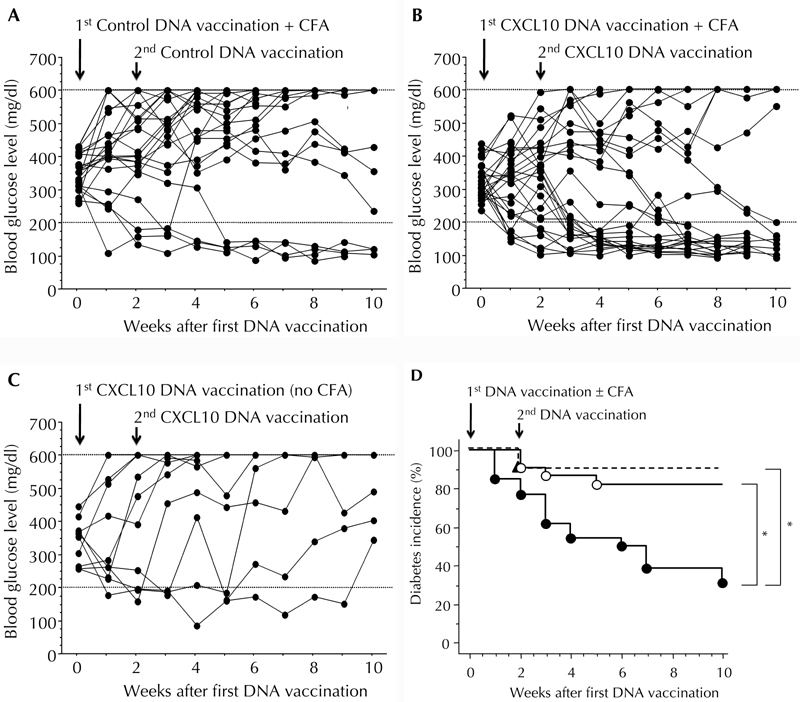
Objective: Complete Freund's Adjuvant (CFA) is known to arrest autoimmune diabetes development in non-obese diabetic (NOD) mice. However, CFA alone cannot induce effective remission in diabetic NOD mice. Previously, we reported that anti-CXC chemokine ligand 10 (CXCL10) antibody can promote beta-cell proliferation in NOD mice. In the present study, we aimed to examine whether anti-CXCL10 plus CFA treatment can effectively reverse autoimmune diabetes development.

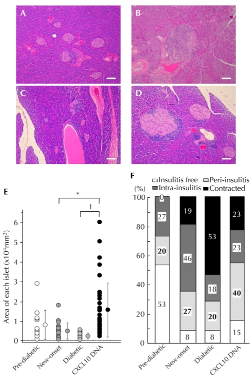
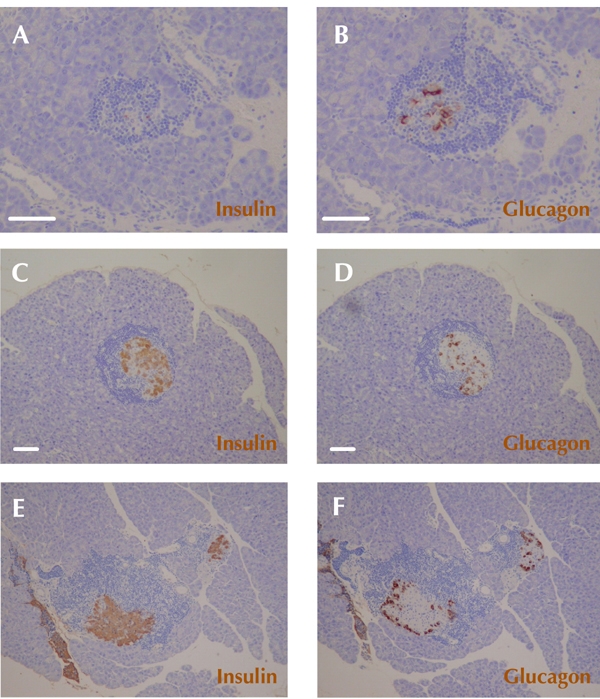
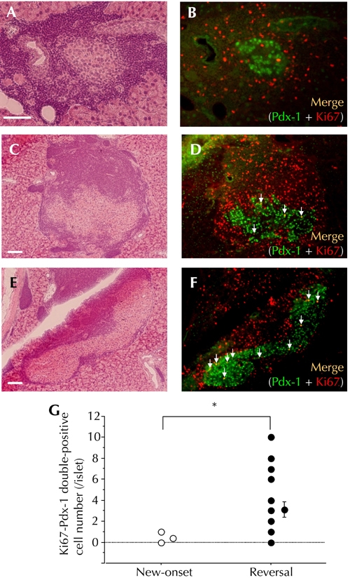
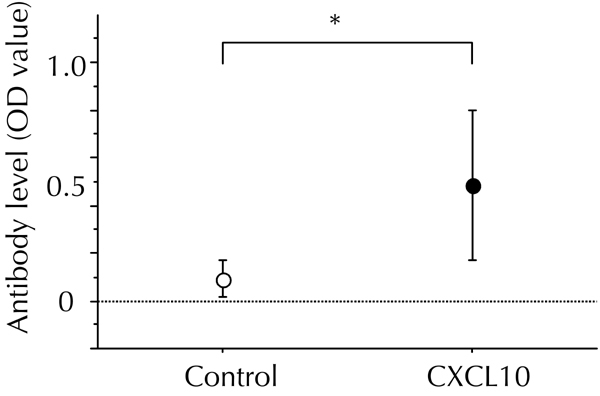
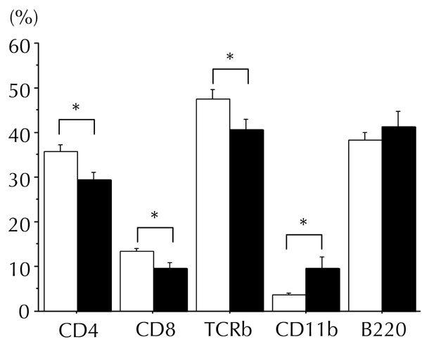
Methods: Systemic supply of anti-CXCL10 antibody by CXCL10 DNA vaccination in combination with CFA injection was performed in new-onset diabetic NOD mice. Remission rate of diabetes, histological characteristics of residual insulitis lesions, residual beta-cell mass, and regulatory T cell population in local pancreas were examined.

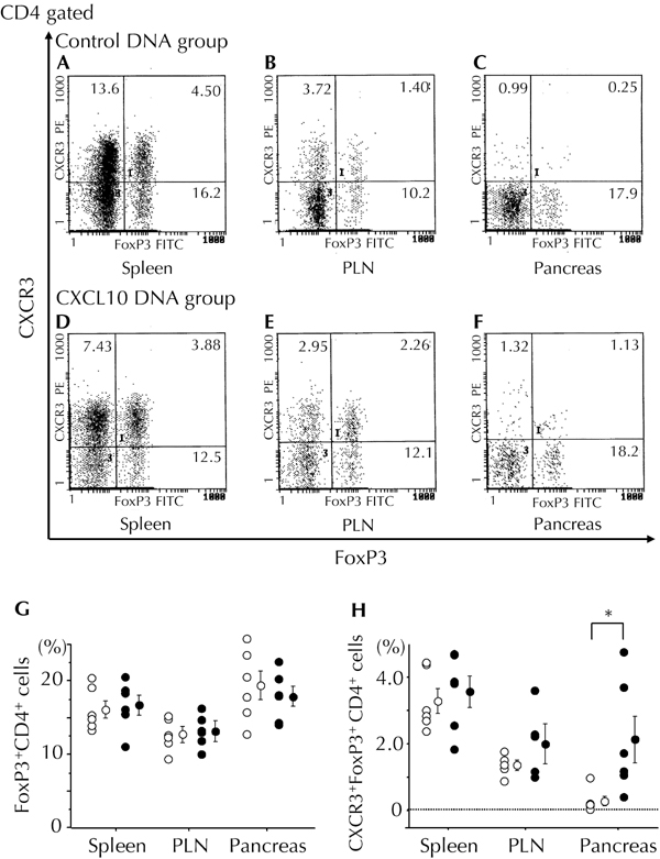
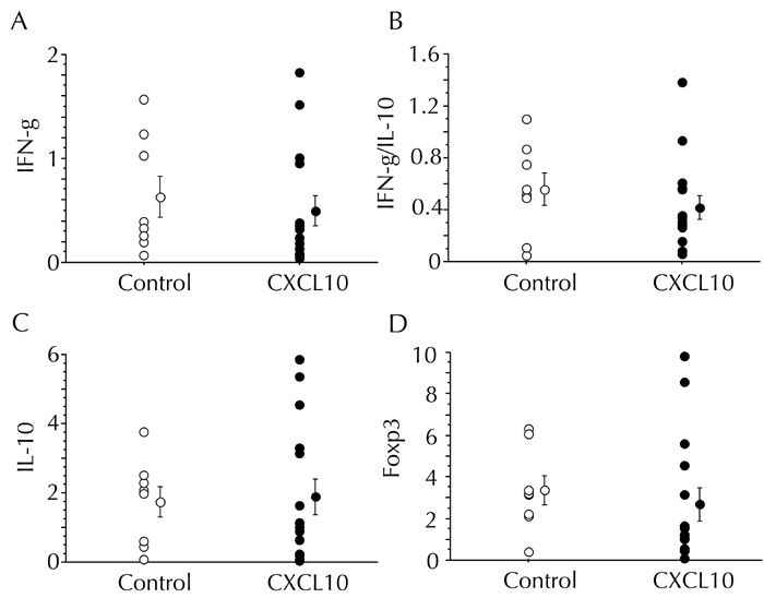
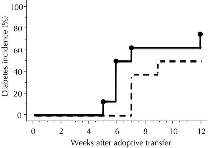
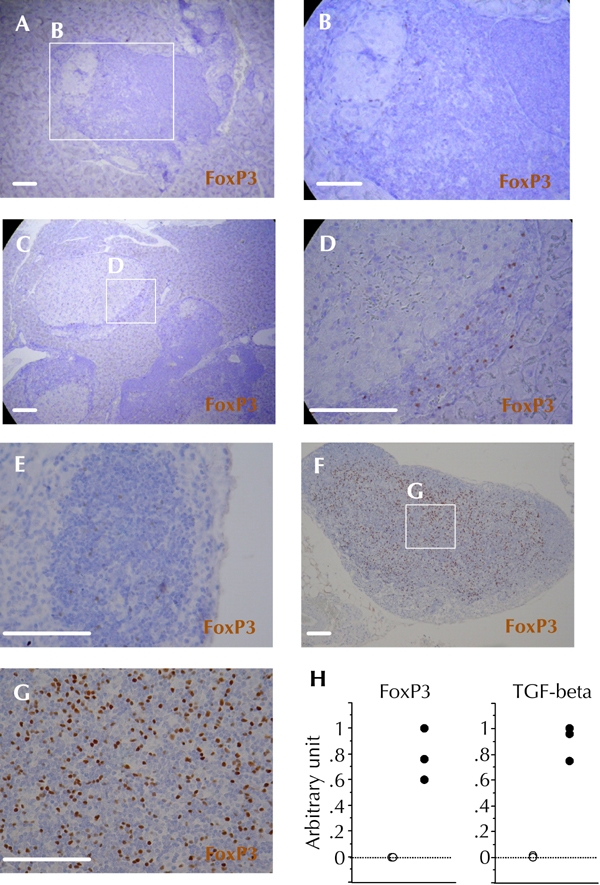
Results: A high frequency of diabetes reversal was observed after combination treatment with anti-CXCL10 plus CFA. In mice showing diabetes reversal, residual beta-cell mass was significantly increased, and some beta-cells were in a proliferative state. Although systemic cytokine profiles were unaffected, the frequency of "hybrid regulatory T cells", i.e. regulatory T cells expressing CXCR3, was significantly increased in local pancreatic lesions. This was possibly associated with the regulation of anti-islet autoimmunity.

Conclusions: Anti-CXCL10 plus appropriate immune adjuvant therapy arrested, and reversed, type 1 diabetes development.


Figures

Figure 1
Figure 1. CXCL10 DNA vaccination in combination with CFA injection effectively reversed hyperglycemia in new-onset diabetic NOD mice
A, B, C: Time courses of blood glucose levels in NOD mice after the indicated treatments. A: Mice treated with control DNA vaccination plus CFA injection (n = 22). B: Mice treated with CXCL10 DNA vaccination plus CFA injection (n = 26). C: Mice treated with CXCL10 DNA vaccination alone (n = 10). D: Kaplan-Meier plot of cumulative incidence in diabetes remission after the indicated treatments. Closed circles and solid lines: Mice treated with CXCL10 DNA vaccination plus CFA injection. Open circles and solid lines: Mice treated with control DNA vaccination plus CFA injection. Closed triangles and dotted lines: Mice treated with CXCL10 DNA vaccination alone. * p < 0.05 by the log-rank test.
Figure 2
Figure 2. Increased residual islet areas in CXCL10 DNA group with reversal of hyperglycemia
A-D: HE staining. Representative islet lesions in pre-diabetic NOD mice (A), new-onset diabetic NOD mice (B), established diabetic NOD mice in control DNA group (C), and CXCL10 DNA group with reversal of hyperglycemia (D). E: Islet areas in the four groups were measured and plotted. Residual islet areas were larger in mice with reversal of hyperglycemia from the CXCL10 DNA group. F: Degrees of insulitis in the four groups. Each group consisted of 8 mice, and at least 30 islets from each mouse were observed by two independent observers. More than half of the residual islets in the CXCL10 DNA group with reversal of hyperglycemia showed peri-insulitis or no insulitis (D). * p < 0.05, p < 0.01 by Mann-Whitney U test. Scale bars: 80 μm.
Figure 3
Figure 3. Alpha-cells scattered within residual islets in the group of CXCL10 DNA-treated mice with reversal of hyperglycemia
Immunohistochemical staining of insulin (A, C, E) and glucagon (B, D, F) was performed. A and B: Representative insulitis lesions in established diabetic NOD mice in the group of control DNA mice. C and D: Representative insulitis lesions in the group of CXCL10 DNA-treated mice with reversal of hyperglycemia 8 to 10 weeks after first DNA vaccination. Glucagon-positive cells (α-cells) are scattered within remaining islets (D). E and F: Representative typical distribution patterns of α-cells and β-cells in insulitis lesions of a new-onset diabetic NOD mice. Scale bars: 80 μm.
Figure 4
Figure 4. Increased residual β-cells in a proliferative state in the group of CXCL10 DNA-treated mice with reversal of hyperglycemia
HE staining (A, C, E) and immunohistochemical double staining of Pdx-1 and Ki67 (B, D, F) were performed. A and B: Representative insulitis lesion in a new-onset diabetic NOD mouse. C, D, E, and F: Two representative insulitis lesions in a CXCL10 DNA vaccinated mouse with reversal of hyperglycemia at 8 to 10 weeks after first DNA vaccination. White arrows indicate Pdx-1 and Ki67 double-positive islet cells (C, D: intra-insulitis, E, F: peri-insulitis). G: The numbers of Pdx-1 and Ki67 double-positive islet cells per islet were counted and plotted. Open circles: new-onset diabetic NOD mice (n = 6, total of 24 islets). Filled circles: CXCL10 DNA-treated mice with reversal of hyperglycemia (n = 8, total of 24 islets). * p < 0.01 by Mann-Whitney U test. Scale bars: 80 μm.
Figure 5
Figure 5. Increased local hybrid Treg cell population in the pancreas of mice in the CXCL10 DNA group
Treg cell-related populations in the spleen (A and D), pancreatic lymph nodes (B and E), and local pancreatic tissue (C and F) were measured by flow cytometry 4 weeks after first DNA vaccination. Representative flow cytometric data from the control DNA group (A-C) and the CXCL10 DNA group (D-F) are shown. G and H: The ratio of FoxP3+CD4+ cells to CD4+ cells (G) and the ratio of CXCR3+FoxP3+CD4+ (hybrid Treg) cells to CD4+ cells (H) are shown. Open circles: control DNA group (n = 6). Filled circles: CXCL10 DNA group (n = 6). * p < 0.05 by Mann-Whitney U test. PLN: pancreatic lymph node.
Figure A1
Figure A1. Anti-mouse CXCL10 antibody was induced by CXCL10 DNA vaccination
In combination with CFA treatment, the first DNA vaccination with pCAGGS-CXCL10 (CXCL10 DNA vaccination), or pCAGGS control plasmid DNA (control DNA vaccination), was performed in new-onset diabetic NOD mice. Each DNA vaccination was repeated two weeks later. 8 to 10 weeks after the DNA vaccination, a direct ELISA was performed to measure the OD values of anti-mouse CXCL10 antibody in sera. The OD values in the control DNA group (n = 9) and the CXCL10 DNA group (n = 9) were 0.09 ± 0.04 and 0.49 ± 0.10, respectively. * p < 0.05 by Mann-Whitney U test.
Figure A2
Figure A2. Lymphocyte populations in the spleen were affected by CXCL10 DNA vaccination in combination with CFA injection
Populations of splenic lymphocytes (CD4+ cells, CD8+ cells, whole T cells (TCR-β), macrophages (CD11b), and B cells (B220)) were measured by flow cytometry 8 to 10 weeks after first DNA vaccination. Counts of splenic CD4+, CD8+, and whole T cells were slightly reduced in the group of CXCL10 DNA-treated mice with reversal of hyperglycemia (filled bar, n = 7), as compared to the control DNA group (open bar, n = 7). In contrast, splenic macrophage count was increased. Cell populations are presented as the ratio of each cell count to that of gated lymphocytes. * p < 0.05 by Mann-Whitney U test.
Figure A3
Figure A3. Splenic cytokines and FoxP3 mRNA expression levels were not affected by CXCL10 vaccination in combination with CFA injection
The expression levels of IFN-γ, IL-10, and FoxP3 mRNAs in the spleen were measured by semi-quantitative real-time PCR 8 to 10 weeks after first DNA vaccination. The control DNA group (n = 8) and the CXCL10 DNA group (n=12) are indicated by open and filled circles, respectively. A, C, and D: Data are presented in arbitrary units. B: The ratio of IFN-γ to IL-10 mRNA expression levels is presented.
Figure A4
Figure A4. Anti-islet autoimmunity persists in the spleens of mice with reversal of hyperglycemia in the CXCL10 DNA group
Adoptive transfer of whole splenocytes from the CXCL10 DNA group with reversal of hyperglycemia, or from control DNA group without reversal, was performed in female NOD-SCID mice 8 to 10 weeks after the first DNA vaccination. The incidence of diabetes development was investigated. The control DNA group (n = 8) and the CXCL10 DNA group (n = 8) are indicated by dotted and solid lines, respectively. There was no significant difference in diabetes incidence between the two groups, calculated by log-rank test.
Figure A5
Figure A5. FoxP3-positive cells persisted in both insulitis lesions and pancreatic lymph nodes in the group of CXCL10 DNA-treated mice long after reversal of hyperglycemia
Immunohistochemical staining of FoxP3 was performed more than 10 weeks after the first DNA vaccination. A and B: Representative insulitis lesion in the control DNA group without reversal of hyperglycemia. C and D: Representative insulitis lesion in the CXCL10 DNA group with reversal of hyperglycemia. E, F, and G: Representative pancreatic lymph node in the control DNA group without reversal of hyperglycemia (E), and in the CXCL10 DNA group with reversal of hyperglycemia (F and G). H: FoxP3 and TGF-β mRNA expression levels in pancreatic lymph nodes were measured in control DNA group mice without reversal of hyperglycemia (open circles, n = 3), and in CXCL10 DNA group mice with reversal of hyperglycemia (filled circles, n = 3) by semi-quantitative real-time PCR. Scale bars: 80 μm.

References

    1. Atkinson MA, Eisenbarth GS. Type 1 diabetes: new perspectives on disease pathogenesis, and treatment. Lancet. 2001;358:221–229. - PubMed
    1. Kikutani H, Makino S. The murine autoimmune diabetes model: NOD, and related strains. Adv Immunol. 1992;51:285–322. - PubMed
    1. Delovitch TL, Singh B. The nonobese diabetic mouse as a model of autoimmune diabetes: immune dysregulation gets the NOD. Immunity. 1997;7:727–738. - PubMed
    1. Loetscher M, Gerber B, Loetscher P, Jones SA, Piali L, Clark-Lewis I, Baggiolini M, Moser B. Chemokine receptor specific for IP10, and mig: structure, function, and expression in activated T-lymphocytes. J Exp Med. 1996;184(3):963–969. - PMC - PubMed
    1. Shimada A, Morimoto J, Kodama K, Suzuki R, Oikawa Y, Funae O, Kasuga A, Saruta T, Narumi S. Elevated serum IP-10 levels observed in type 1 diabetes. Diabetes Care. 2001;24(3):510–515. - PubMed

Publication types

MeSH terms

LinkOut - more resources