. 2011 Jun;85(11):5406-14.
doi: 10.1128/JVI.02190-10. Epub 2011 Mar 16.

A new Ebola virus nonstructural glycoprotein expressed through RNA editing

Affiliations

A new Ebola virus nonstructural glycoprotein expressed through RNA editing

Masfique Mehedi et al. J Virol. 2011 Jun.

Abstract

Ebola virus (EBOV), an enveloped, single-stranded, negative-sense RNA virus, causes severe hemorrhagic fever in humans and nonhuman primates. The EBOV glycoprotein (GP) gene encodes the nonstructural soluble glycoprotein (sGP) but also produces the transmembrane glycoprotein (GP₁,₂) through transcriptional editing. A third GP gene product, a small soluble glycoprotein (ssGP), has long been postulated to be produced also as a result of transcriptional editing. To identify and characterize the expression of this new EBOV protein, we first analyzed the relative ratio of GP gene-derived transcripts produced during infection in vitro (in Vero E6 cells or Huh7 cells) and in vivo (in mice). The average percentages of transcripts encoding sGP, GP₁,₂, and ssGP were approximately 70, 25, and 5%, respectively, indicating that ssGP transcripts are indeed produced via transcriptional editing. N-terminal sequence similarity with sGP, the absence of distinguishing antibodies, and the abundance of sGP made it difficult to identify ssGP through conventional methodology. Optimized 2-dimensional (2D) gel electrophoresis analyses finally verified the expression and secretion of ssGP in tissue culture during EBOV infection. Biochemical analysis of recombinant ssGP characterized this protein as a disulfide-linked homodimer that was exclusively N glycosylated. In conclusion, we have identified and characterized a new EBOV nonstructural glycoprotein, which is expressed as a result of transcriptional editing of the GP gene. While ssGP appears to share similar structural properties with sGP, it does not appear to have the same anti-inflammatory function on endothelial cells as sGP.


Figures
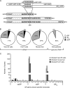
Fig. 1.
Fig. 1.
Ebola virus glycoprotein gene RNA editing results in multiple gene products. (A) Organization of the Ebola virus glycoprotein gene. (Top) Putative open reading frames (ORFs) for the different GP gene products (sGP, GP1,2, and ssGP). (Bottom) The primary structures of glycoprotein gene products are shown in an alignment of the primary amino acid sequences of sGP, GP1,2, and ssGP. All three proteins share the first 295 aa, including the signal peptide (aa 1 to 32), but differ in their carboxy-terminal portions. (B) Detection of ssGP transcripts in vitro and in vivo. Vero E6 and Huh7 cells were infected with ZEBOV, and mice were infected with MA-ZEBOV. RNA was extracted from infected cell cultures, virus particles, and mouse liver. The editing site region for mRNA and vRNA was amplified (330-bp fragment), cloned, and sequenced as described in Materials and Methods. In total, we analyzed 224 clones from Vero E6 cells, 132 from Huh7 cells, 260 from mouse liver, and 109 from viral particles. (C) Specificity of transcriptional editing. The vast majority of clones from mRNA (97%) contained either 7 (sGP), 8 (GP1,2), or 9 (ssGP) adenosine (A) residues. The remaining 3% of clones analyzed contained multiple adenosine residues in the editing site, which would lead to the incorporation of additional lysine residues in the primary amino acid sequence. With the rare exception of a single adenosine deletion (in clones encoding ssGP), no deletions of adenosine residues were found in the editing site.
Fig. 2.
Fig. 2.
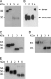
Structure and biochemical properties of ssGP. (A) ssGP is a homodimer. Vero E6 cells were transfected with the appropriate expression plasmids, and supernatants were collected from transfected cells after 72 h. The proteins were separated by reducing or nonreducing SDS-PAGE and were detected by immunoblotting using MAb 42/3.7 (dilution, 1:10,000). Lane 1, r.ssGP (nonreducing); lane 2; r.sGP (nonreducing); lane 3, r.ssGP (reducing); lane 4, r.sGP (reducing). (B) Cysteine at position 53 is responsible for dimerization. Site-directed mutagenesis was performed at amino acid position 53 (cysteine to glycine), and the resulting plasmid was transfected into 293T cells. Proteins were detected by immunoblotting with a peroxidase-conjugated anti-HA antibody (dilution, 1:10,000). Lane 1, r.ssGP Cys53Gly mutant (reducing); lane 2, r.ssGP (reducing); lane 3, r.ssGP Cys53Gly mutant (nonreducing); lane 4, r.ssGP (nonreducing). (C) ssGP contains N-linked carbohydrates. Vero E6 cells were transfected with the appropriate expression plasmids, and supernatants were collected from transfected cells after 72 h. The proteins were treated with PNGase F, separated by reducing SDS-PAGE, and detected by immunoblotting using MAb 42/3.7 (dilution, 1:10,000). Lane 1, untreated r.ssGP; lane 2, untreated r.sGP; lane 3, PNGase F-treated r.ssGP; lane 4, PNGase F-treated r.sGP. (D) ssGP does not contain high-mannose-type N-linked carbohydrates. 293T cells were transfected with the appropriate plasmid expressing HA-tagged r.ssGP, and HA-tagged r.ssGP was purified from the supernatants 72 h posttransfection (see Materials and Methods). HA-tagged r.ssGP was treated with Endo H and/or PNGase F and was analyzed by reducing SDS-PAGE followed by immunoblotting using peroxidase-conjugated anti-HA (dilution, 1:10,000). Lane 1, untreated r.ssGP; lane 2, Endo H-treated r.ssGP; lane 3, r.ssGP treated with Endo H and PNGase F; lane 4, r.ssGP treated with PNGase F. (E) ssGP does not contain O-linked carbohydrates. HA-tagged r.ssGP treated with different exoglycosidases, O-glycanase, N-glycanase, or combinations of these glycosidases was analyzed as described in Materials and Methods. Lane 1, untreated r.ssGP; lane 2, r.ssGP treated with the exoglycosidases [sialidase A, β(1–4)-galactosidase, and β-N-acetylglucosaminidase]; lane 3, r.ssGP treated with the exoglycosidases and O-glycanase; lane 4, r.ssGP treated with the exoglycosidases, O-glycanase, and N-glycanase; lane 5, r.ssGP treated with N-glycanase.
Fig. 3.
Fig. 3.
Expression of ssGP during Ebola virus infection. Vero E6 cells were infected with ZEBOV at an MOI of 1. Supernatants were collected 4 days postinfection and were treated with PNGase F. Subsequently, proteins were separated using different electrophoresis systems and were detected by immunoblotting using MAb 42/3.7 at a 1/10,000 dilution. For controls, Vero E6 cells were transfected with the appropriate plasmids expressing r.sGP and r.ssGP. Supernatants were collected 72 h posttransfection and were analyzed as described above. (A) Reducing 15% SDS-PAGE. Lane 1, r.ssGP treated with PNGase F; lane 2, r.sGP treated with PNGase F; lane 3, supernatant from ZEBOV-infected Vero E6 cells treated with PNGase F. (B) 2D gel electrophoresis. (Left) r.ssGP and r.sGP treated with PNGase F. (Right) Supernatant from ZEBOV-infected Vero E6 cells treated with PNGase F.
Fig. 4.
Fig. 4.
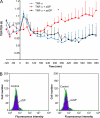
Functional analysis of ssGP. (A) Endothelial barrier function rescue assay. Human umbilical vein endothelial cells were equilibrated for 2 h to generate a baseline TER. TNF-α (1 ng/ml) with or without ssGP or sGP (30 μg/ml) was added to the medium as indicated, and the chambers were monitored by impedance spectroscopy. There was a significant difference (P < 0.05) between the TER of cells treated with TNF-α alone and that of cells treated with TNF-α and sGP by 330 min (*), while there was no significant difference between cells treated with TNF-α alone and cells treated with TNF-α and ssGP. (B) Neutrophil binding assay. Purified human neutrophils were incubated with ssGP or sGP (20 μg/ml) and were subsequently stained with anti-HA-PE and analyzed by FACS. The lack of a shift in fluorescence intensity between untreated and treated neutrophils indicates that neither sGP nor ssGP binds to the surfaces of human neutrophils.

Similar articles

Cited by

References

    1. Barrientos L. G., Martin A. M., Rollin P. E., Sanchez A. 2004. Disulfide bond assignment of the Ebola virus secreted glycoprotein SGP. Biochem. Biophys. Res. Commun. 323:696–702 - PubMed
    1. Barrientos L. G., Martin A. M., Wohlhueter R. M., Rollin P. E. 2007. Secreted glycoprotein from live Zaire ebolavirus-infected cultures: preparation, structural and biophysical characterization, and thermodynamic stability. J. Infect. Dis. 196(Suppl. 2):S220–S231 - PubMed
    1. Bourhis J. M., Canard B., Longhi S. 2006. Structural disorder within the replicative complex of measles virus: functional implications. Virology 344:94–110 - PubMed
    1. Bray M., Davis K., Geisbert T., Schmaljohn C., Huggins J. 1998. A mouse model for evaluation of prophylaxis and therapy of Ebola hemorrhagic fever. J. Infect. Dis. 178:651–661 - PubMed
    1. Casey J. L., Bergmann K. F., Brown T. L., Gerin J. L. 1992. Structural requirements for RNA editing in hepatitis delta virus: evidence for a uridine-to-cytidine editing mechanism. Proc. Natl. Acad. Sci. U. S. A. 89:7149–7153 - PMC - PubMed

Publication types

Substances