. 2011 May;85(10):5091-104.
doi: 10.1128/JVI.02565-10. Epub 2011 Mar 16.

Reverse genetic analysis of Ourmiaviruses reveals the nucleolar localization of the coat protein in Nicotiana benthamiana and unusual requirements for virion formation

Affiliations

Reverse genetic analysis of Ourmiaviruses reveals the nucleolar localization of the coat protein in Nicotiana benthamiana and unusual requirements for virion formation

Giulia Crivelli et al. J Virol. 2011 May.

Abstract

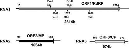
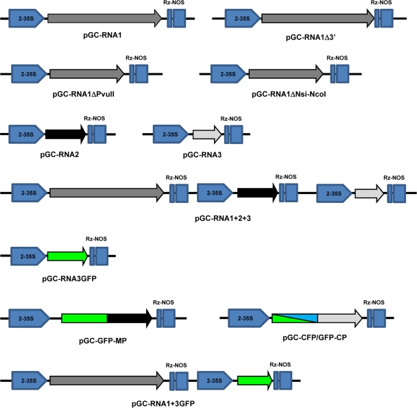
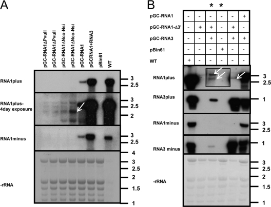
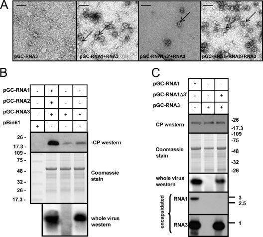
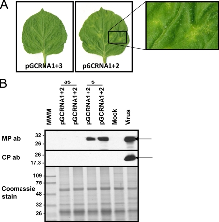
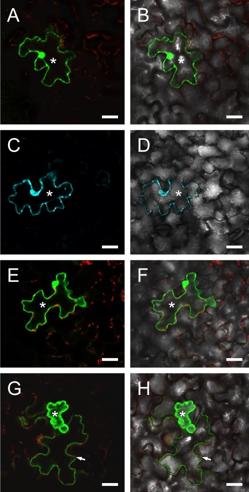
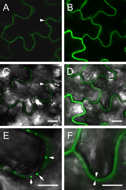
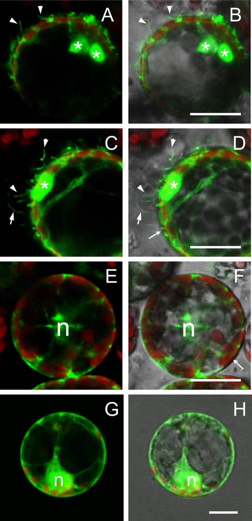
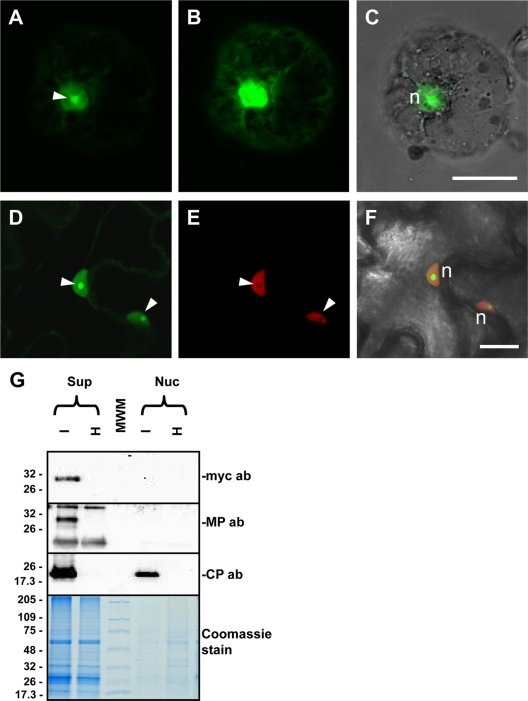
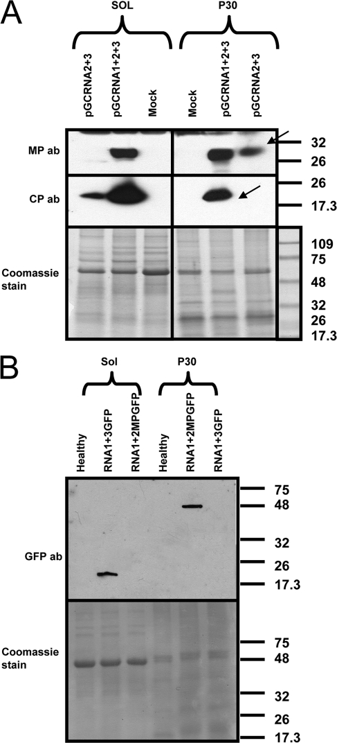
Ourmia melon virus (OuMV) is the type member of the genus Ourmiavirus. These viruses have a trisegmented genome, each part of which encodes a single protein. Ourmiaviruses share a distant similarity with other plant viruses only in their movement proteins (MP), whereas their RNA-dependent RNA polymerase (RdRP) shares features only with fungal viruses of the family Narnaviridae. Thus, ourmiaviruses are in a unique phylogenetic position among existing plant viruses. Here, we developed an agroinoculation system to launch infection in Nicotiana benthamiana plants. Using different combinations of the three segments, we demonstrated that RNA1 is necessary and sufficient for cis-acting replication in the agroinfiltrated area. RNA2 and RNA3, encoding the putative movement protein and the coat protein (CP), respectively, are both necessary for successful systemic infection of N. benthamiana. The CP is dispensable for long-distance transport of the virus through vascular tissues, but its absence prevents efficient systemic infection at the exit sites. Virion formation occurred only when the CP was translated from replication-derived RNA3. Transient expression of a green fluorescent protein-MP (GFP-MP) fusion via agroinfiltration showed that the MP is present in cytoplasmic connections across plant cell walls; in protoplasts the GFP-MP fusion stimulates the formation of tubular protrusions. Expression through agroinfiltration of a GFP-CP fusion displays most of the fluorescence inside the nucleus and within the nucleolus in particular. Nuclear localization of the CP was also confirmed through Western blot analysis of purified nuclei. The significance of several unusual properties of OuMV for replication, virion assembly, and movement is discussed in relation to other positive-strand RNA viruses.


Figures

Fig. 1.
Fig. 1.
OuMV genome organization. Each encoded ORF is indicated by an arrow. The most important restriction sites used in the mutagenesis process are shown.
Fig. 2.
Fig. 2.
Schematic representation of constructs built and used in this study. Each clone has the same pJL89-derived backbone, where double StuI/SmaI restriction digestion allows insertion of blunt-end PCR fragments derived from RT-PCR of full-length genomic segments between the double 35S promoter (2-35S) and the hepatitis delta virus ribozyme (Rz) followed by a Nos terminator. Arrow directions indicate the orientation of the viral transcripts obtained for each of the described clones. The arrows indicate each ORF encoded by the genome.
Fig. 3.
Fig. 3.
RNA1 encodes a fully competent cis- and trans-acting replicase. (A) Northern blot analysis of total RNA extracted from agroinfiltrated areas 3 days postinfiltration (dpi). The top panel shows viral plus-strand RNA evaluated with a 2-h exposure to the X-ray film; the middle panel shows the same blot exposed for 4 days to evaluate accumulation of the deleted RNA1 transcript (arrow); the bottom panels show the minus-strand RNA accumulation with a 4-day exposure and the rRNA, respectively. Each agroinfiltrated clone is detailed at the top of each lane and graphically described in Fig. 2. (B) Northern blot analysis of RNA extracted at 3 dpi with a combination of agroclones specified above the panels (except for the samples corresponding to the lanes marked by asterisks, which were instead processed 2 dpi). From top to bottom, each of the four panels shows plus-strand RNA1, plus-strand RNA3, minus-strand RNA1, and minus-strand RNA3. The bottom panel shows rRNA stained with methylene blue on the membrane. The inset replaces the original 2-day exposure with a 6-day exposure. The arrows indicate the two primary transcripts from pGC-RNA1Δ3′ (ribozyme processed and full-length). The RNA ladder bands are those from BrightStar Biotinylated RNA Millennium Markers (Ambion), and the sizes are expressed in kb.
Fig. 4.
Fig. 4.
Virion formation analysis in agroinfiltrated areas at 3 days postagroinfiltration (dpi) with the combination of agroclones described in Fig. 2. (A) Electron micrograph of negatively stained ISEM preparations of partially purified virus preparation from agroinfiltrated leaves. The arrows indicate single viral particles. Bar, 50 nm. (B) The top panel shows Western blotting of SDS-PAGE-separated protein extracts from the agroinfiltrated area to detect OuMV coat protein (CP); the middle panel shows Coomassie staining for total protein extracts separated on SDS-PAGE, and the bottom panel shows a Western blot for the CP after partial virus purification and whole-virus agarose gel separation to detect virion presence. Combinations of the binary plasmids used in each agro-suspension treatment are specified at the top of the blots. (C) The three top panels are the same as in panel B; the two bottom panels show the results of Northern blot analysis for OuMV RNA1 and RNA3, respectively, extracted from partially purified virus preparations. The RNA ladder bands are those from BrightStar Biotinylated RNA Millennium Markers (Ambion), and the sizes are expressed in kb. Thermo Scientific Pierce Blue Prestained MW Marker (Pierce) was used for Western blot analysis, and the sizes of the bands are expressed in kDa.
Fig. 5.
Fig. 5.
Limited necrotic areas in upper uninoculated leaves when the plant is agroinfiltrated with a mix of RNA1- and RNA2-expressing clones. (A) Symptoms on upper uninoculated leaves at 12 days postinfiltration (dpi) with a combination of agrobacterium clones. The inset to the right shows an enlargement of the localized symptomatic area. (B) Western blotting for the presence of the OuMV MP (upper panel) and CP (middle panel). Samples were collected at 15 dpi in symptomatic (s) and asymptomatic (as) areas of an upper uninoculated leaf. Mock, inoculation with empty pBin61; virus, infection by mechanical inoculation of sap from N. benthamiana infected with OuMV VE9 isolate. The bottom panel shows Coomassie staining of the proteins loaded in the SDS-PAGE gel. Thermo Scientific Pierce Blue Prestained MW Marker (Pierce) is the molecular weight marker (MWM), and the sizes of the bands are expressed in thousands. The arrows indicate the position of the MP and CP bands in the Western blot. ab, antibody.
Fig. 6.
Fig. 6.
Cell-to-cell movement of GFP-MP fusion expressed in epidermal cells. Agro-suspensions precisely diluted at an optical density at 600 nm of 0.5 were agroinfiltrated in 1/10 serial dilutions until single-cell transfection was obtained. Only fully developed N. benthamiana leaves were agroinfiltrated. All pictures correspond to the 10−3 dilution. Fluorescence images (A, C, E, and G) are overlaid with the respective bright-field images (B, D, F, and H). (A and B) Localization of pBinGFP-CP fluorescence limited to the transfected epidermal cell (asterisk). The weak red signal (A and B, E and F, and G and H) derives from excitation of chloroplast autofluorescence in the mesophyll. Analogous single-cell localization (asterisk) of pBin-CFP (C and D) fluorescence is visible as well as for pBin-GFP (E and F). When pBin-GFP-MP was agroinfiltrated (G and H), fluorescence leaked from the transfected cell (asterisk) to the neighboring cells (arrowhead). Bar, 20 μm.
Fig. 7.
Fig. 7.
Subcellular localization of GFP-MP versus GFP in N. benthamiana leaf epidermal cells, observed 3 days postagroinfiltration. Images show localization of pGC-GFP-MP (A, C, and E) and pGC-RNA3-GFP (B, D, and F) in agroinfiltrated leaves. The fluorescence images in panels A and B are overlaid with the corresponding bright-field images in panels C and D; only the overlaid pictures are shown for the higher magnification images in panels E and F. In panels A and C the punctate pattern of MP-GFP fluorescence and its association with the cell wall are visible (arrowhead); panel E shows a particularly evident pattern (arrowheads). Localization of free GFP (B and D) to the cytoplasm clearly identifies the unlabeled cell wall (F) as a dark line separating the fluorescent cytoplasms of adjacent cells (arrowheads). Bars, 10 μm (A to D) and 5 μm (E and F).
Fig. 8.
Fig. 8.
Subcellular localization of GFP-MP fusion in N. benthamiana protoplasts. (A, C, E, and G) Fluorescent signals of GFP (green) and chloroplasts (red). (B, D, F, and H) The same pictures overlaid with the corresponding bright-field images. Protoplasts transfected with the pGC-GFP-MP construct are shown in panels A to D. Besides the large aggregates visible in the cytoplasm (asterisks), GFP-MP fluorescence highlights numerous tubular protrusions of the protoplast plasma membrane. Such protrusions were never observed in protoplasts expressing the cytosolic GFP construct from the pGC-RNA3-GFP construct (E and F) or the endoplasmic reticulum-associated GFP-ER (G and H). n, nucleus. Bar, 20 μm.
Fig. 9.
Fig. 9.
Nucleolar localization of GFP-CP fusion protein. Images show subcellular localization of GFP-CP fusion in N. benthamiana protoplasts (A to C) and epidermal cells (D to F) and Western blotting for CP and MP localization on nuclear extracts (G). (A) In protoplasts, GFP-CP shows the strongest fluorescent intensity in the nucleolus (arrowhead) although clear labeling is present in the whole nucleus. (B) A weaker signal is visible in the cytoplasm after the gain of the photo multiplier tube was increased. (C) Overlay of the image in panel A with the corresponding bright-field image to visualize cell boundaries. An analogous strong labeling of the nucleolus is visible in leaf epidermal cells. In this case, the GFP-CP construct was agroinfiltrated in transgenic plants expressing H2B-RFP. (D) GFP fluorescence, where strong nucleolar labeling is evident (arrowheads). (E) Nucleoli are also visible as dark unlabeled spots in the corresponding H2B-RFP image, due to the exclusion of H2B from the nucleolus. (F) Both fluorescent signals overlaid with the bright-field image of the same cells. n, nucleus. Bar 20 μm. (G) Western blot showing the presence of OuMV CP in a purified nuclear extract from OuMV-infected N. benthamiana plants. No MP is present in the extract, as demonstrated by the lack of labeling with both anti-MP and anti-Myc antibodies (ab) since a Myc-MP fusion was agroinfiltrated and used for the OuMV infection. Thermo Scientific Pierce Blue Prestained MW Marker (Pierce) is the molecular weight marker (MWM), and the sizes of the bands are expressed in thousands.
Fig. 10.
Fig. 10.
Distribution of OuMV CP and MP in subcellular fractions during active viral infection or coexpressed through agroinfiltration in the absence of RNA1. (A) Western blot of proteins from virus infection (pG-CRNA1+2+3) and coagroinfiltration of RNA2 and RNA3 (pGC-RNA2+3) in the absence of RNA1. Samples were processed at 4 dpi. Arrows indicate the specific CP and MP bands. ab, antibody. (B) Control for proper separation between cytosolic and noncytosolic fraction. GFP was expressed from pBin-GFP through agroinfiltration, and samples for Western blotting were processed at 3 dpi. GFP-MP is a fusion of GFP with the movement protein obtained from agroinfiltration of pBin-GFP-MP and processed at 3 dpi. Sol, cytosolic fraction; P30, noncytosolic, membranous fraction; mock, pBin61-infiltrated leaf; ab, antibody. Bottom panels show Coomassie staining of a replicate of the gel used for Western blotting. Thermo Scientific Pierce Blue Prestained MW Marker (Pierce) is the molecular weight marker (MWM), and the sizes of the bands are expressed in thousands.

References

    1. Accotto G. P., Milne R. G. 2008. Ourmiavirus, p. 500–501In Mahy B. W., Van Regenmortel M. H. V.(ed.), Encyclopedia of virology. Elsevier, London, United Kingdom
    1. Agranovsky A. A., et al. 1998. Beet yellows closterovirus HSP70-like protein mediates the cell-to-cell movement of a potexvirus transport-deficient mutant and a hordeivirus-based chimeric virus. J. Gen. Virol. 79:889–895 - PubMed
    1. Aiton M. M., Lennon A. M., Roberts I. M., Harrison B. D. 1988. Two new cassava viruses from Africa, p. 43.In Proceedings of the 5th International Congress of Plant Pathology, Kyoto, Japan Phytopathological Society of Japan, Tokyo, Japan
    1. Annamalai P., Rao A. L. N. 2006. Packaging of brome mosaic virus subgenomic RNA is functionally coupled to replication-dependent transcription and translation of coat protein. J. Virol. 80:10096–10108 - PMC - PubMed
    1. Annamalai P., Rao A. L. N. 2005. Replication-independent expression of genome components and capsid protein of brome mosaic virus in planta: a functional role for viral replicase in RNA packaging. Virology 338:96–111 - PubMed

Publication types

MeSH terms

Substances