. 2011 May;85(10):4993-5002.
doi: 10.1128/JVI.02265-10. Epub 2011 Mar 16.

Activities of ICP0 involved in the reversal of silencing of quiescent herpes simplex virus 1

Affiliations

Activities of ICP0 involved in the reversal of silencing of quiescent herpes simplex virus 1

Michael W Ferenczy et al. J Virol. 2011 May.

Abstract

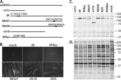
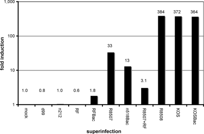
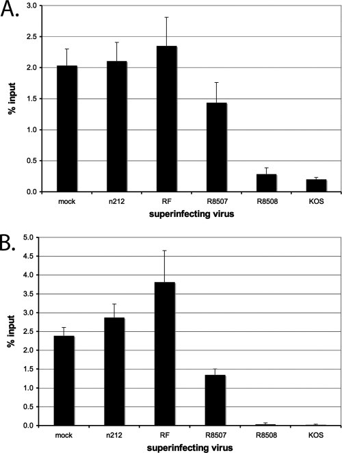
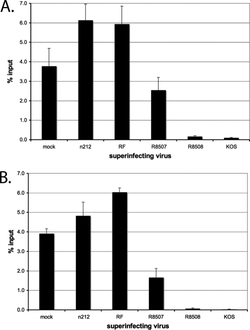
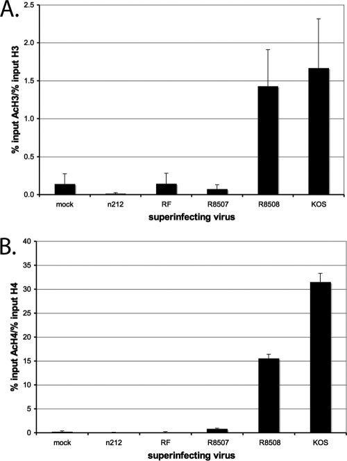
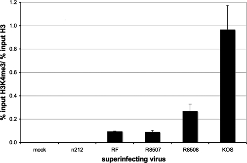
ICP0 is a transcriptional activating protein required for the efficient replication and reactivation of latent herpes simplex virus 1 (HSV-1). Multiple regions of ICP0 contribute its activity, the most prominent of which appears to be the RING finger, which confers E3 ubiquitin ligase activity. A region in the C terminus of ICP0 has also been implicated in several activities, including the disruption of a cellular repressor complex, REST/CoREST/HDAC1/2/LSD1. We used quiescent infection of MRC-5 cells with a virus that does not express immediate-early proteins, followed by superinfection with various viral mutants to quantify the ability of ICP0 variants to reactivate gene expression and alter chromatin structure. Superinfection with wild-type virus resulted in a 400-fold increase in expression from the previously quiescent d109 genome, the removal of heterochromatin and histones from the viral genome, and an increase in histone marks associated with activated transcription. RING finger mutants were unable to reactivate transcription or remove heterochromatin from d109, while mutants that are unable to bind CoREST activate gene expression from quiescent d109, albeit to a lesser degree than the wild-type virus. One such mutant, R8507, resulted in the partial removal of heterochromatin. Infection with R8507 did not result in the hyperacetylation of H3 and H4. The results demonstrate that (i) consistent with previous findings, the RING finger domain of ICP0 is required for the activation of quiescent genomes, (ii) the RF domain is also crucial for the ultimate removal of repressive chromatin, (iii) activities or interactions specified by the carboxy-terminal region of ICP0 significantly contribute to activation, and (iv) while the effects of the R8507 on chromatin are consistent with a role for REST/CoREST/HDAC1/2/LSD1 in the repression of quiescent genomes, the mutation may also affect other activities involved in derepression.


Figures

Fig. 1.
Fig. 1.
Viruses used in theses studies. Panel A shows a representation of the ICP0 alleles in the indicated mutants (wt, wild type). Some of these mutants were used to superinfect MRC-5 cells that were infected for 1 week with d109 (MOI = 10 PFU/cell). GFP expression was assessed 24 h later by fluorescence microcopy (B). One-week confluent cultures of MRC-5 cells were also infected with wild-type and ICP0 mutant viruses for 4 h and labeled with [35S]methionine, and SDS-PAGE samples were prepared as described in Materials and Methods. SDS peptides were electrophoretically separated, transferred to a nitrocellulose membrane, and analyzed by Western blotting for ICP0 (C). The membrane was then exposed to X-ray film for visualization of all of the infected cell polypeptides (D). The values to the right are molecular sizes in kilodaltons.
Fig. 2.
Fig. 2.
Relative abundance of GFP mRNA in d109-infected MRC-5 cells after superinfection or coinfection. Infections, RNA isolation, cDNA preparation, and RT-PCR were performed as described in Materials and Methods. Quiescent infection by d109 was established for 1 week and followed by superinfection with the indicated mutant(s). Each value indicates the fold induction (over mock superinfection) of GFP mRNA due to the indicated superinfections.
Fig. 3.
Fig. 3.
Repressive chromatin modifications associated with the HCMV promoter of d109 in MRC-5 cells after superinfection. Quiescent infection by d109 was established for 1 week. ChIP with antibodies to HP1γ (A) and H3K9me3 (B), followed by RT-PCR, was performed as described in Materials and Methods. Shown are the percentages of total genomes bound after superinfection with the indicated virus at 4 h after superinfection with the indicated virus. Error bars represent standard error of the mean from multiple experiments.
Fig. 4.
Fig. 4.
Binding of histones H3 and H4 to the HCMV promoter of d109 in MRC-5 cells after superinfection. ChIP with antibodies to histones H3 and H4 and RT-PCR were performed as described in the legend to Fig. 2 to determine the percentages of genomes bound by histones H3 (A) and H4 (B). Graphs show the percentage of total genomes bound at 4 h after superinfection with the indicated virus. Error bars represents the standard error of the mean from multiple experiments.
Fig. 5.
Fig. 5.
Binding of hyperacetylated histone H3 (AcH3) and hyperacetylated H4 (AcH4) to the HCMV promoter of d109 in MRC-5 cells after superinfection. ChIP with an antibody to AcH3 (A) or AcH4 (B) and RT-PCR were performed as described in the legend to Fig. 2 to determine the percentage of d109 genomes bound by AcH3 or AcH4 at 4 h after superinfection with the indicated virus. AcH3 was normalized to the amount of histone H3 (A) and AcH4 was normalized to the amount of histone H4 (B) on the genome by dividing the percentage of acetylated histone by the percentage of total histone (Fig. 3). Error bars represent standard deviations from multiple experiments.
Fig. 6.
Fig. 6.
Binding of histone H3 dimethyl lysine 4 (H3K4me2) to the HCMV promoter of d109 in MRC-5 cells after superinfection. ChIP with an antibody to H3K4me2 and RT-PCR were performed as described in the legend to Fig. 2 to determine the percentage of d109 genomes bound by H3K4me2 at 4 h after superinfection with the indicated virus. H3K4me2 was normalized to the amount of histone H3 on the genome by dividing the percentage of acetylated histone by the percentage of total histone (Fig. 3). Error bars represent standard deviations from multiple experiments.

References

    1. Bannister A. J., et al. 2001. Selective recognition of methylated lysine 9 on histone H3 by the HP1 chromo domain. Nature 410:120–124 - PubMed
    1. Barlow P. N., Luisi B., Milner A., Elliott M., Everett R. 1994. Structure of the C3HC4 domain by 1H-nuclear magnetic resonance spectroscopy. A new structural class of zinc-finger. J. Mol. Biol. 237:201–211 - PubMed
    1. Birnboim H. C., Doly J. 1979. A rapid alkaline extraction procedure for screening recombinant plasmid DNA. Nucleic Acids Res. 7:1513–1523 - PMC - PubMed
    1. Boutell C., Sadis S., Everett R. D. 2002. Herpes simplex virus type 1 immediate-early protein ICP0 and its isolated RING finger domain act as ubiquitin E3 ligases in vitro. J. Virol. 76:841–850 - PMC - PubMed
    1. Cai W., et al. 1993. The herpes simplex virus type 1 regulatory protein ICP0 enhances virus replication during acute infection and reactivation from latency. J. Virol. 67:7501–7512 - PMC - PubMed

Publication types

Substances

LinkOut - more resources