Systemic dissemination of H5N1 influenza A viruses in ferrets and hamsters after direct intragastric inoculation
- PMID: 21411541
- PMCID: PMC3126210
- DOI: 10.1128/JVI.00148-11
Systemic dissemination of H5N1 influenza A viruses in ferrets and hamsters after direct intragastric inoculation
Abstract
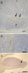
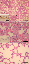
Although oral exposure to H5N1 highly pathogenic avian influenza viruses is a risk factor for infection in humans, it is unclear how oral exposure to these virus results in lethal respiratory infections. To address this issue, we inoculated ferrets and hamsters with two highly pathogenic H5N1 strains. These viruses, inoculated directly into the stomach, were isolated from the large intestine and the mesenteric lymph nodes within 1 day of inoculation and subsequently spread to multiple tissues, including lung, liver, and brain. Histopathologic analysis of ferrets infected with virus via direct intragastric inoculation revealed lymph folliculitis in the digestive tract and mesenteric lymph nodes and focal interstitial pneumonia. Comparable results were obtained with the hamster model. We conclude that, in mammals, ingested H5N1 influenza viruses can disseminate to nondigestive organs, possibly through the lymphatic system of the gastrointestinal tract.
Figures
Similar articles
-
Pathogenesis of H5N1 influenza virus infections in mice and ferret models differs according to respiratory tract or digestive system exposure.J Infect Dis. 2009 Mar 1;199(5):717-25. doi: 10.1086/596740. J Infect Dis. 2009. PMID: 19210164
-
Pathogenesis of Influenza A/H5N1 virus infection in ferrets differs between intranasal and intratracheal routes of inoculation.Am J Pathol. 2011 Jul;179(1):30-6. doi: 10.1016/j.ajpath.2011.03.026. Epub 2011 May 5. Am J Pathol. 2011. PMID: 21640972 Free PMC article.
-
Effect of an asparagine-to-serine mutation at position 294 in neuraminidase on the pathogenicity of highly pathogenic H5N1 influenza A virus.J Virol. 2011 May;85(10):4667-72. doi: 10.1128/JVI.00047-11. Epub 2011 Mar 2. J Virol. 2011. PMID: 21367898 Free PMC article.
-
[Clue to the molecular mechanism of virulence of highly pathogenic H5N1 avian influenza viruses isolated in 2004].Uirusu. 2005 Jun;55(1):55-61. doi: 10.2222/jsv.55.55. Uirusu. 2005. PMID: 16308530 Review. Japanese.
-
The current state of H5N1 vaccines and the use of the ferret model for influenza therapeutic and prophylactic development.J Infect Dev Ctries. 2012 Jun 15;6(6):465-9. doi: 10.3855/jidc.2666. J Infect Dev Ctries. 2012. PMID: 22706187 Review.
Cited by
-
H5N1 pathogenesis studies in mammalian models.Virus Res. 2013 Dec 5;178(1):168-85. doi: 10.1016/j.virusres.2013.02.003. Epub 2013 Feb 28. Virus Res. 2013. PMID: 23458998 Free PMC article. Review.
-
Marked endotheliotropism of highly pathogenic avian influenza virus H5N1 following intestinal inoculation in cats.J Virol. 2012 Jan;86(2):1158-65. doi: 10.1128/JVI.06375-11. Epub 2011 Nov 16. J Virol. 2012. PMID: 22090101 Free PMC article.
-
Comparative Pathology of Animal Models for Influenza A Virus Infection.Pathogens. 2023 Dec 29;13(1):35. doi: 10.3390/pathogens13010035. Pathogens. 2023. PMID: 38251342 Free PMC article. Review.
-
Pathogenesis and Transmission of Novel Highly Pathogenic Avian Influenza H5N2 and H5N8 Viruses in Ferrets and Mice.J Virol. 2015 Oct;89(20):10286-93. doi: 10.1128/JVI.01438-15. Epub 2015 Jul 29. J Virol. 2015. PMID: 26223637 Free PMC article.
-
The multibasic cleavage site in H5N1 virus is critical for systemic spread along the olfactory and hematogenous routes in ferrets.J Virol. 2012 Apr;86(7):3975-84. doi: 10.1128/JVI.06828-11. Epub 2012 Jan 25. J Virol. 2012. PMID: 22278228 Free PMC article.
References
-
- Abdel-Ghafar A. N., et al. 2008. Update on avian influenza A (H5N1) virus infection in humans. N. Engl. J. Med. 358:261–273 - PubMed
-
- Ali M. J., Teh C. Z., Jennings R., Potter C. W. 1982. Transmissibility of influenza viruses in hamsters. Arch. Virol. 72:187–197 - PubMed
-
- Cavriani G., et al. 2005. Lymphatic system as a path underlying the spread of lung and gut injury after intestinal ischemia/reperfusion in rats. Shock 23:330–336 - PubMed
-
- Enserink M., Kaiser J. 2004. Virology. Avian flu finds new mammal hosts. Science 305:1385. - PubMed
Publication types
MeSH terms
LinkOut - more resources
Full Text Sources
Medical

