. 2011 Mar 16;31(11):4280-9.
doi: 10.1523/JNEUROSCI.5310-10.2011.

Aversive stimuli alter ventral tegmental area dopamine neuron activity via a common action in the ventral hippocampus

Affiliations

Aversive stimuli alter ventral tegmental area dopamine neuron activity via a common action in the ventral hippocampus

Ornella Valenti et al. J Neurosci. .

Abstract

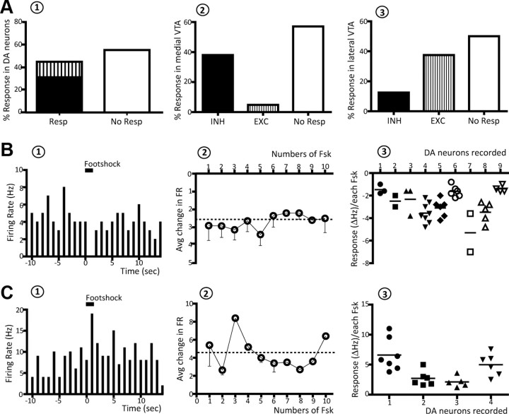
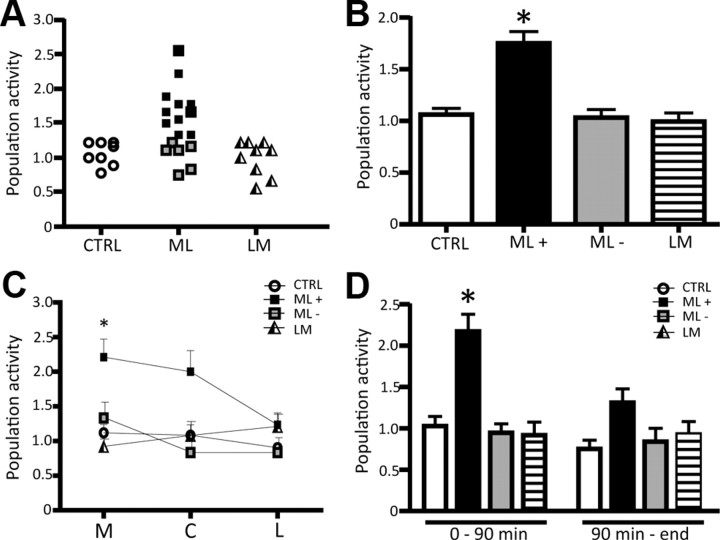
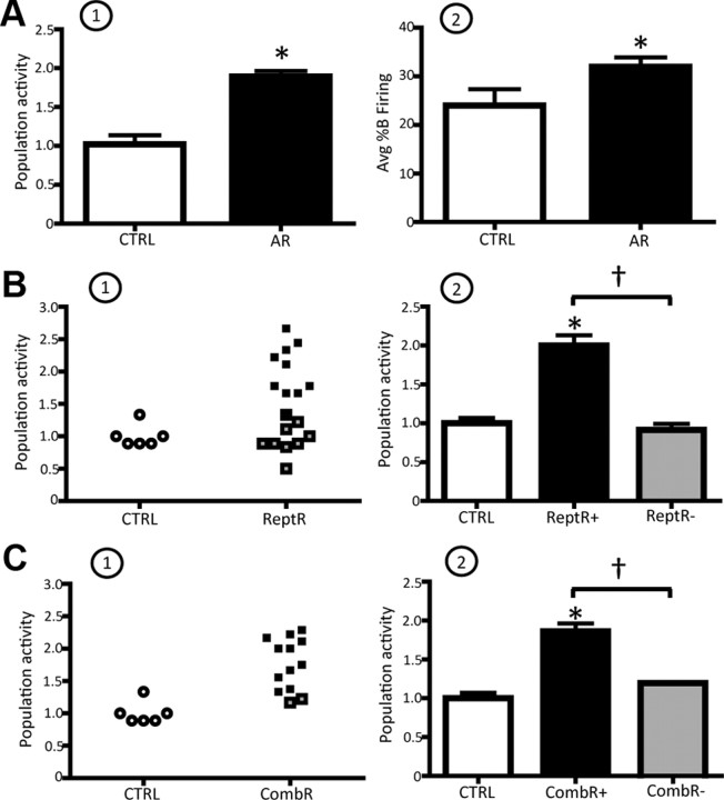
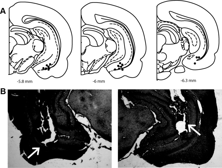
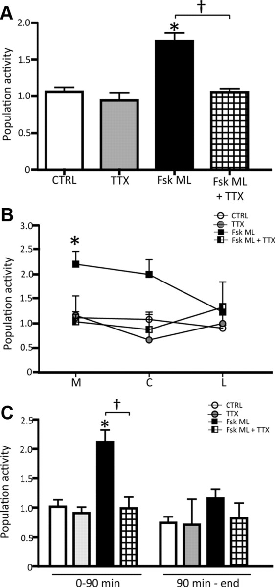
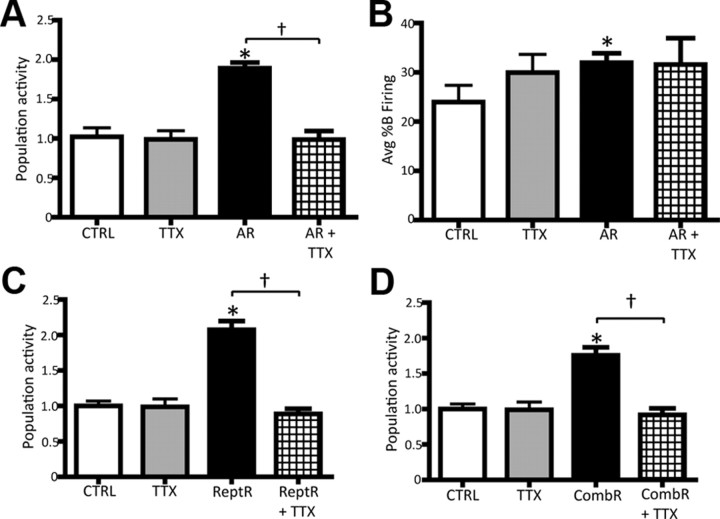
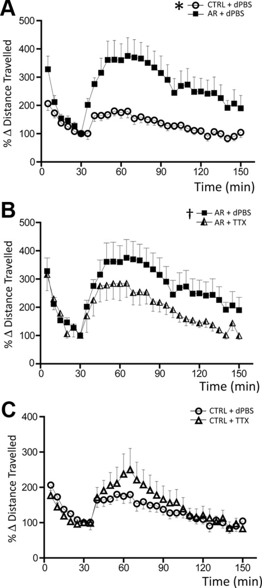
Stress is a physiological, adaptive response to changes in the environment, but can also lead to pathological alterations, such as relapse in psychiatric disorders and drug abuse. Evidence demonstrates that the dopamine (DA) system plays a role in stress; however, the nature of the effects of sustained stressors on DA neuron physiology has not been adequately addressed. By using a combined electrophysiological, immunohistochemical and behavioral approach, we examined the response of ventral tegmental area DA neurons in rats to acute as well as repeated stressful events using noxious (footshock) and psychological (restraint) stress. We found that aversive stimuli induced a pronounced activation of the DA system both electrophysiologically (population activity; i.e., number of DA neurons firing spontaneously) and behaviorally (response to psychostimulants). Moreover, infusion of TTX into the ventral hippocampus (vHPC) reversed both behavioral and electrophysiological effects of stress, indicating that the hyperdopaminergic condition associated with stress is driven by hyperactivity within the vHPC. Therefore, the stress-induced activation of the DA system may underlie the propensity of stress to exacerbate psychotic disorders or predispose an individual to drug-seeking behavior. Furthermore, the vHPC represents a critical link between context-dependent DA sensitization, stress-induced potentiation of amphetamine responsivity, and the increase in DA associated with stressors.


Figures

Figure 1.
Figure 1.
Repeated noxious stimuli induced both inhibition and excitation of VTA DA neurons. A, Effects of footshock on identified DA neuron activity. A1, Approximately 45% of DA neurons tested responded to footshock, and displayed either inhibition (bottom of first bar, black) or excitation (top of first bar, vertical lines). A2, A3, Neurons located in the medial portion of the VTA (A2) were predominantly inhibited whereas DA neurons located in the lateral portions of the VTA (A3) more often showed excitation. B1, Footshock produced an inhibition in firing of this representative neuron; footshock is delivered at t = 0 and the duration of the stimulus indicated by the horizontal line. B2, Average change in FR of footshock-inhibited neurons; each point represents the average (average) response of all neurons recorded, and is repeated during all 10 footshocks. The magnitude of the response was uniform across each footshock; dashed horizontal line represents average mean change in FR. B3, Individual neurons showed variable amplitudes of inhibition to footshocks. In this plot different symbols are used for each neuron recorded (x-axis), and within the same neuron each data point represents a response (expressed as ΔHz) to a given footshock; note that not all of the 10 footshocks evoked a response. Horizontal lines indicate the average response for each neuron. As can be observed, although individual neurons showed marked differences in amplitude of response, within a given neuron the responses were more tightly clustered in amplitude. C, Same as B, but for neurons that were excited by footshock.
Figure 2.
Figure 2.
Repeated footshock (Fsk) stimulation causes a selective increase in population activity of medial VTA DA neurons. A, Distribution of DA neuron population activity (number of spontaneously active DA neurons encountered per electrode track) for control (CTRL) rats (open circles), and group ML (black squares, DA activated; gray circles, DA nonactivated) and LM (triangles) of rats receiving footshock (each symbol represents the response in a single rat). B, The ML group of rats was segregated into two groups: those rats showing a significant increase in population activity (ML+; black bar) and those that were DA nonactivated (ML−; gray bar). Recordings made in LM (horizontal line bar) rats did not reveal differences in population activity compared to control (white bar). C, When the regions are analyzed separately, a significant increase in population activity is observed in the medial (M), but not in the central (C) or lateral (L) VTA. D, The increase in population activity was also limited in duration, in that significant differences were only observed within 90 min following delivery of the footshock. Significant difference between Fsk rats and CTRL (one-way RM ANOVA); for all values, *p < 0.001.
Figure 3.
Figure 3.
Restraint stress potently activates VTA DA neurons. A1, A2, AR induced a pronounced increase in the average population activity (A1) and in the average percentage burst firing (A2) of the VTA DA neurons recorded. B1, Distribution of population activity observed in control (CTRL; open circles) versus ReptR; DA-activated rat data are shown in black; DA-nonactivated data are shown in gray (each symbol represents the response in a single rat). B2, ReptR induced a twofold increase in population activity in DA-activated rats, but not in the non-DA-activated group. C, Effect of CombR (repeated plus acute) on VTA DA neuron population activity. Color code: white, controls; black, DA activated following restraint; gray, DA nonactivated. *Significant difference between CTRL and restraint rats; significant difference in repeated restraint in DA-activated vs -nonactivated rats. Population activity, CTRL vs AR: one-way ANOVA, p < 0.001; CTRL vs ReptR: one-way RM ANOVA, p < 0.001; CTRL vs CombR: one-way RM ANOVA, p < 0.001. Avg %B firing, CTRL vs AR: one-way ANOVA, p = 0.043.
Figure 4.
Figure 4.
Acute restraint stress induced a selective increase in c-fos expression in the medial PFC and vHPC. A, Acute restraint causes a significant increase in c-fos expression in the vHPC, shown as an increase in c-fos staining (black dots). A1, Schematic of a coronal slice illustrating the region under study; A2, c-fos expression in controls; A3, c-fos expression following acute restraint stress. B, Acute restraint fails to activate c-fos in the dHPC. C, Acute restraint causes a substantial activation of c-fos expression also in the medial PFC (mPFC).
Figure 5.
Figure 5.
Location of cannulae placements for vHPC inactivation in electrophysiology and behavioral experiments. A, Representative localization of cannulae placements (filled dots) in rat vHPC coronal sections for local infusion of 1 μm TTX (0.5 μl) (electrophysiology experiments). B, Arrows indicate location of bilateral cannula placements for TTX infusion in representative behavioral experiment in the left and right vHPC, respectively.
Figure 6.
Figure 6.
The vHPC is required for the footshock-induced increase in VTA DA neuron population activity. A–C, Local infusion of TTX into the vHPC either prevented or reversed (see Results) the footshock-induced increase in DA neuron population activity (A) that predominantly occurred in the medial VTA (B) and within the first 90 min following repeated footshocks (C). Color code for bar diagram and symbols: white, controls; gray, control + TTX; black, footshock; grid, footshock + TTX. Significant difference between footshock (group ML, DA activated only) and CTRL (one-way RM ANOVA), *p < 0.001; significant difference between footshock (Fsk) ML and Fsk ML + TTX (one-way RM ANOVA), p < 0.001.
Figure 7.
Figure 7.
Inactivation of the vHPC reversed the restraint-induced increase in DA neuron population activity. A, Population activity in control (CTRL) and AR rats; TTX administered into the vHPC following restraint reversed the increase in population activity in the AR rats but did not affect controls. B, In contrast, inactivation of the vHPC did not affect the AR-induced increase in percentage burst firing. C, The effects of ReptR stress on DA neuron population activity and its reversal by infusion of TTX into the vHPC. D, Effect of CombR stress on population activity and its reversal by infusion of TTX. Color code for bar diagram and symbols: white, controls; gray, control + TTX; black, footshock; grid, footshock + TTX. *Significant difference between restraint and CTRL; significant difference between restraint and restraint + TTX; AR vs AR+ TTX: one-way ANOVA, p < 0.001; ReptR vs ReptR + TTX: one-way RM ANOVA, p < 0.001; CombR vs CombR + TTX: one-way RM ANOVA, p = 0.001.
Figure 8.
Figure 8.
Inactivation of the vHPC reverses the acute restraint-induced potentiation of amphetamine-induced locomotion. A, Time course of amphetamine-induced increase in locomotor activity, comparing AR (black squares) and control (CTRL; open circles). In acute restraint animals, administration of 1.5 mg/kg amphetamine caused a substantially greater increase in locomotor activity compared with nonstressed controls. B, Infusion of TTX into the vHPC reversed the stress-induced augmentation of amphetamine-induced locomotion in the AR rats (triangles, AR followed by infusion of TTX). C, Inactivation of the vHPC did not affect the locomotor response to amphetamine in control rats (open triangles; CTRL + TTX). Significant difference between CTRL + dPBS and AR + dPBS (two-way ANOVA), *p < 0.001; significant difference between AR + dPBS and AR + TTX (two-way ANOVA), p = 0.004.

Similar articles

Cited by

References

    1. Abercrombie ED, Keefe KA, DiFrischia DS, Zigmond MJ. Differential effect of stress on in vivo dopamine release in striatum, nucleus accumbens, and medial frontal cortex. J Neurochem. 1989;52:1655–1658. - PubMed
    1. Anstrom KK, Woodward DJ. Restraint increases dopaminergic burst firing in awake rats. Neuropsychopharmacology. 2005;30:1832–1840. - PubMed
    1. Antelman SM, Eichler AJ, Black CA, Kocan D. Interchangeability of stress and amphetamine in sensitization. Science. 1980;207:329–331. - PubMed
    1. Bhatnagar S, Sun LM, Raber J, Maren S, Julius D, Dallman MF. Changes in anxiety-related behaviors and hypothalamic-pituitary-adrenal activity in mice lacking the 5-HT-3A receptor. Physiol Behav. 2004;81:545–555. - PubMed
    1. Bouton ME, Bolles RC. Role of conditioned contextual stimuli in reinstatement of extinguished fear. J Exp Psychol Anim Behav Process. 1979;5:368–378. - PubMed

Publication types

MeSH terms