Comparative Study
. 2011 May;59(5):504-17.
doi: 10.1369/0022155411400875. Epub 2011 Mar 16.

Histochemical localization of caldesmon in the CNS and ganglia of the mouse

Affiliations
Comparative Study

Histochemical localization of caldesmon in the CNS and ganglia of the mouse

Christoph N Köhler. J Histochem Cytochem. 2011 May.

Abstract

The author has recently reported the distribution of the cytoskeleton-associated protein caldesmon in spleen and lymph nodes detected with different antibodies against caldesmon (J Histochem Cytochem 58:183-193, 2010). Here the author reports the distribution of caldesmon in the CNS and ganglia of the mouse using the same antibodies. Western blot analysis of mouse brain and spinal cord showed the preponderance of l-caldesmon and suggested at least two l-caldesmon isoforms in the brain. Immunostaining revealed the predominant reactivity of smooth muscle cells and cells resembling pericytes of many large and small blood vessels, ependymocytes, and secretory cells of the pineal gland and pituitary gland. Neuronal perikarya and neuropil in general displayed no or weak immunoreactivity, but there was stronger labeling of neuronal perikarya in dorsal root and trigeminal ganglia. In the brain, staining of the neuropil was stronger in the molecular layers of the dentate gyrus and cerebellum. Results show that caldesmon is expressed in many different cell types in the CNS and ganglia, consistent with the notion that l-caldesmon is ubiquitously expressed, but it appears most concentrated in smooth muscle cells, pericytes, epithelial cells, secretory cells, and neuronal perikarya in dorsal root and trigeminal ganglia.


Conflict of interest statement

The author(s) declared no potential conflicts of interest with respect to the authorship and/or publication of this article.

Figures
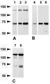
Figure 1.
Figure 1.
Western blots of brain (lanes 1, 2, 4, 5) and spinal cord (lanes 3, 6) after 6% SDS-PAGE stained with the caldesmon rabbit polyclonal antibody (A) and clone 8/L-Caldesmon (B). Lanes 1 and 4, 10 µg per lane loaded; lanes 2, 3, 5, 6, 20 µg per lane loaded. The rabbit polyclonal and clone 8/L-Caldesmon recognize two closely spaced bands at ~71/72 kDa clearly visible after reduced protein load. The rabbit polyclonal antibody stains a band at 130 kDa corresponding to the molecular weight of h-caldesmon in addition. (C) Western blots of uterus (2.5 µg per lane loaded) show that the rabbit polyclonal antibody (lane 7) and clone 8/L-Caldesmon (lane 8) recognize h-caldesmon as well as two bands within the molecular weight range of l-caldesmon.
Figure 2.
Figure 2.
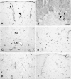
Overview of caldesmon immunoreactivity of the mouse brain. (A) Four percent paraformaldehyde (PFA)–fixed, sagittal paraffin section of a mouse brain stained with the caldesmon rabbit polyclonal antibody after microwave pretreatment in citrate buffer. Larger blood vessels (large arrows), ependymal cells lining the ventricles and aqueduct (3V, third ventricle; 4V, fourth ventricle; Aq, aqueduct), ependymal cells of the choroid plexus (arrowheads), and the pineal (Pin) and pituitary (Pit) gland display strong immunostaining clearly recognizable at low magnification, but medium-sized (small arrows point to examples) and small blood vessels are also stained. If compared to unstained white matter (cc, corpus callosum; df, dorsal fornix; py, pyramidal tract), there is weak immunoreactivity of the neuropil, which is stronger in the molecular layer of the cerebellum. (B) Immunoreactivity is absent after replacement of the caldesmon rabbit polyclonal antibody by an irrelevant rabbit polyclonal antibody against Aβ42. Color development time was the same for both sections. Scale bar = 2 mm.
Figure 3.
Figure 3.
Caldesmon immunoreactivity of blood vessels in different regions of the brain. (A) Four percent paraformaldehyde (PFA)–fixed paraffin section of the cortex stained with clone 8/L-Caldesmon after microwave pretreatment in EDTA buffer. The meninges and blood vessels are immunoreactive. Cell bodies located at the outer surface of the vessel wall, reminiscent of pericytes (arrowheads point to examples), and fine processes are stained. (B) Four percent PFA-fixed paraffin section of the cerebellum stained with the rabbit polyclonal antibody after microwave pretreatment in citrate buffer. Blood vessels are immunoreactive (arrowheads). Perikarya and major dendrites of Purkinje cells (arrow) are unstained. There is also moderate staining of the molecular layer. (C) Four percent PFA-fixed paraffin section of the hippocampus stained with the rabbit polyclonal antibody after microwave pretreatment in citrate buffer showing immunoreactive blood vessels in the stratum radiatum and lacunosum moleculare layer. (D) Four percent PFA-fixed paraffin section of the superior colliculus stained with clone 8/L-Caldesmon after microwave pretreatment in EDTA buffer showing a dense net of immunoreactive small blood vessels. (E) Four percent PFA-fixed paraffin section of the amygdala stained with the rabbit polyclonal antibody after microwave pretreatment in citrate buffer showing immunoreactive blood vessels. (F) Four percent PFA-fixed cryosection of the cortex stained with the goat (N-19) antibody showing immunoreactive blood vessels. LMol, lacunosum moleculare layer; Rad, stratum radiatum. Scale bar (A, C, D, E, F) = 50 µm. Scale bar (B) = 20 µm.
Figure 4.
Figure 4.
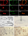
Caldesmon immunoreactivity of smooth muscle cells, pericytes, endothelial cells, and astrocytes. (A–C) Confocal microscopy analysis at 0.5 µm optical thickness of meningeal blood vessels, 4% paraformaldehyde (PFA)–fixed paraffin section. Caldesmon is located in smooth muscle cells. There is only little co-location of caldesmon and the endothelium marker GLUT-1. (D–F) Confocal microscopy analysis at 0.5 µm optical thickness of small cortical blood vessels, 4% PFA-fixed paraffin section. Caldesmon is located in a cell resembling a pericyte (the nucleus is marked by an asterisk in D). There is no co-location of caldesmon and the endothelium marker GLUT-1. (G–I) Confocal microscopy analysis at 0.5 µm optical thickness of small cortical blood vessels, 4% PFA-fixed paraffin section. The rabbit polyclonal antibody and clone 8/L-Caldesmon label the same cells (nuclei are marked by asterisks in G). (J, K) Consecutive 4% PFA-fixed paraffin sections of a medium-sized blood vessel in the brainstem. Cells discontinuously located on the outer surface of the vessel wall (arrows point to examples), reminiscent of pericytes, are stained with the rabbit polyclonal antibody (J) and clone 8/L-Caldesmon (K); the asterisks mark the lumen of the blood vessel. (L) Four percent PFA-fixed paraffin section of the hippocampus. At low magnification, the molecular layer of the dentate gyrus displays moderate and patchy caldesmon immunoreactivity. Note weak immunoreactivity of the oriens layer and stratum radiatum, the staining of blood vessels, but the absence of significant staining of neuronal perikarya, most obviously in the pyramidal and granule cell layers. (M) A section temporarily cover-slipped after staining with the rabbit polyclonal antibody showing the molecular layer of the dentate gyrus at higher magnification. Unstained nuclei and stained coarse processes are visible in the center of the stronger immunoreactive regions (delineated by arrows) resembling astrocytes stained including their fine processes. The arrowhead points to a stronger immunoreactive cell resembling a pericyte. (N) Consecutive staining of the same section for glial fibrillary acidic protein (GFAP) confirms that the moderate caldesmon-immunoreactive cells are astrocytes. The cell resembling a pericyte is not double stained. GrDG, granular layer of dentate gyrus; LMol, lacunosum moleculare layer; Mol, molecular layer of dentate gyrus; Or, oriens layer; Py, pyramidal cell layer; Rad, stratum radiatum. Scale bar: A–C = 20 µm; D–F = 10 µm; G–I = 10 µm; J, K = 50 µm; L = 200 µm; M, N = 50 µm.
Figure 5.
Figure 5.
Caldesmon immunoreactivity of ependymal cells and secretory cells. Consecutive 4% paraformaldehyde (PFA)–fixed paraffin sections stained with the caldesmon rabbit polyclonal antibody (A, D, G) or clone 8/L-Caldesmon (B, E, H) after microwave pretreatment in citrate buffer and comparison with results from in situ hybridization as shown in the Allen Mouse Brain Atlas (C, F) (Allen Mouse Brain Atlas [Internet]. Seattle (WA): Allen Institute for Brain Science. 2009. Available from: http://mouse.brain-map.org). (A–C) Secretory cells of the pineal gland are immunoreactive with the rabbit polyclonal antibody or clone 8/L-Caldesmon, consistent with expression of caldesmon as shown in the Allen Mouse Brain Atlas. (D–F) Sagittal sections of the subcommissural organ show immunostaining of ependymocytes; caldesmon immunoreactivity obtained with the rabbit polyclonal antibody or clone 8/L-Caldesmon is located at their ventricular pole and lateral aspect. Results from the Allen Mouse Brain Atlas show expression in the same cells. Arrows point to immunoreactive ependymal cells lining the third ventricle. (G, H) Mouse pituitary gland consisting of pars distalis (D), pars intermedia (I), and pars nervosa (N). The rabbit polyclonal antibody and clone 8/L-Caldesmon label elongated cells in the pars nervosa. There is also strong staining of epithelial cells lining the cleft of Rathke’s pouch (arrowheads). The pars distalis of the adenohypophysis displays strong immunoreactivity of stellate cells and slender elongated processes lining the sinus as well as immunoreactivity of secretory cells with the rabbit polyclonal antibody. Clone 8/L-Caldesmon stains the same cells but secretory cells weaker. Scale bar: A, B = 100 µm; C = 105 µm; D, E = 50 µm; F = 105 µm; G, H = 100 µm.
Figure 6.
Figure 6.
Caldesmon immunoreactivity of mouse spinal cord and dorsal root ganglia (DRGs). Four percent paraformaldehyde (PFA)–fixed paraffin sections of mouse spinal cord and ganglia stained with the caldesmon rabbit polyclonal antibody (A, C) or clone 8/L-Caldesmon (D). (A) Thoracic spinal cord displays moderately stained gray matter, stronger stained ependymal cells lining the central canal (arrow in A), blood vessels (arrowheads), and meninges. Some neuronal perikarya within the gray matter display moderate but distinct immunoreactivity. (B) Staining is absent from a nearby section when the primary antibody was omitted. (C, D) Neurons in the DRG are moderately stained with the rabbit polyclonal antibody and clone 8/L-Caldesmon. Satellite cells (insert in D) and blood vessels (arrowheads) display stronger immunoreactivity. Scale bar: A, B = 200 µm; C, D = 50 µm.

Similar articles

Cited by

References

    1. Abd-el-Basset EM, Ahmed I, Fedoroff S. 1991. Actin and actin-binding proteins in differentiating astroglia in tissue culture. J Neurosci Res. 30:1–17 - PubMed
    1. Agassandian C, Plantier M, Fattoum A, Represa A, der Terrossian E. 2000. Subcellular distribution of calponin and caldesmon in rat hippocampus. Brain Res. 887:444–449 - PubMed
    1. Agassandian K, Cassell MD. 2008. Co-localization of caldesmon and calponin with cortical afferents, metabotropic glutamate and neurotrophic receptors in the lateral and central nuclei of the amygdala. Brain Res. 21:39–55 - PMC - PubMed
    1. Alexanian AR, Bamburg JR, Hidaka H, Mornet D. 2001. Calcium-dependent regulation of interactions of caldesmon with calcium-binding proteins found in growth cones of chick forebrain neurons. Cell Mol Neurobiol. 21:437–451 - PMC - PubMed
    1. Allt G, Lawrenson JG. 2001. Pericytes: cell biology and pathology. Cells Tissues Organs. 169:1–11 - PubMed

Publication types

Substances