. 2011 Apr 15;186(8):4994-5003.
doi: 10.4049/jimmunol.1003010. Epub 2011 Mar 16.

A unique hybrid renal mononuclear phagocyte activation phenotype in murine systemic lupus erythematosus nephritis

Affiliations

A unique hybrid renal mononuclear phagocyte activation phenotype in murine systemic lupus erythematosus nephritis

Ramalingam Bethunaickan et al. J Immunol. .

Abstract

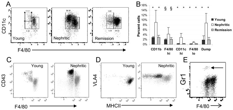
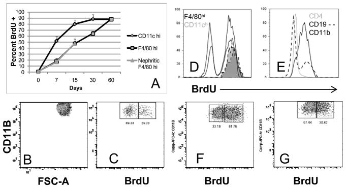
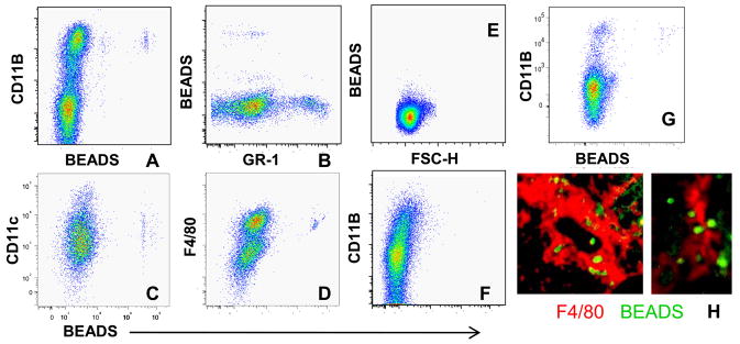
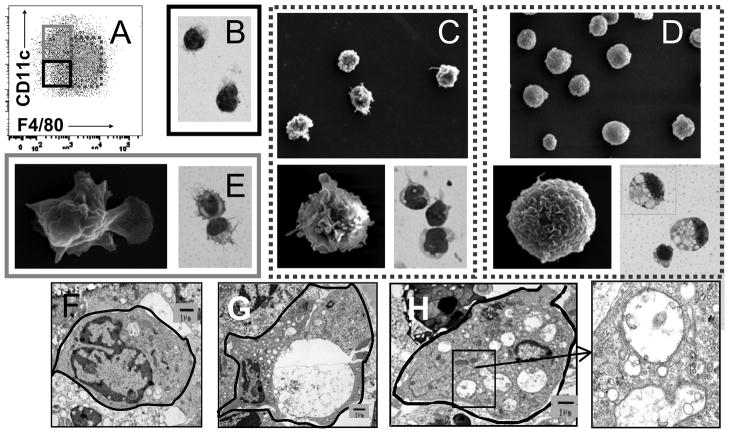
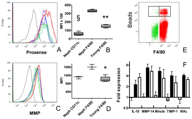
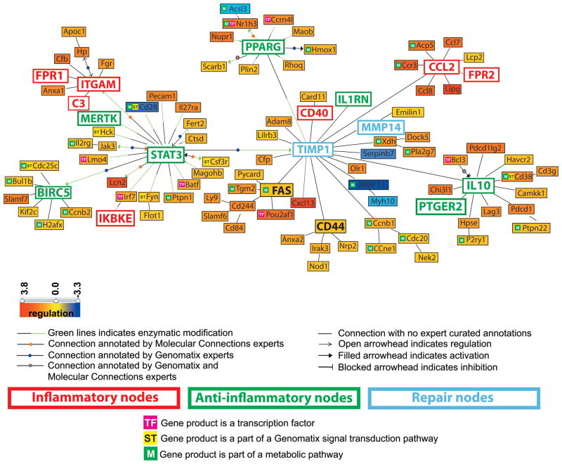
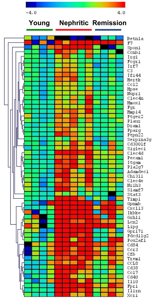
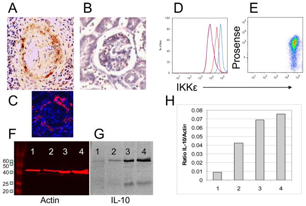
Renal infiltration with mononuclear cells is associated with poor prognosis in systemic lupus erythematosus. A renal macrophage/dendritic cell signature is associated with the onset of nephritis in NZB/W mice, and immune-modulating therapies can reverse this signature and the associated renal damage despite ongoing immune complex deposition. In nephritic NZB/W mice, renal F4/80(hi)/CD11c(int) macrophages are located throughout the interstitium, whereas F4/80(lo)/CD11c(hi) dendritic cells accumulate in perivascular lymphoid aggregates. We show here that F4/80(hi)/CD11c(int) renal macrophages have a Gr1(lo)/Ly6C(lo)/VLA4(lo)/MHCII(hi)/CD43(lo)/CD62L(lo) phenotype different from that described for inflammatory macrophages. At nephritis onset, F4/80(hi)/CD11c(int) cells upregulate cell surface CD11b, acquire cathepsin and matrix metalloproteinase activity, and accumulate large numbers of autophagocytic vacuoles; these changes reverse after the induction of remission. Latex bead labeling of peripheral blood Gr1(lo) monocytes indicates that these are the source of F4/80(hi)/CD11c(int) macrophages. CD11c(hi)/MHCII(lo) dendritic cells are found in the kidneys only after proteinuria onset, turnover rapidly, and disappear rapidly after remission induction. Gene expression profiling of the F4/80(hi)/CD11c(int) population displays increased expression of proinflammatory, regulatory, and tissue repair/degradation-associated genes at nephritis onset that reverses with remission induction. Our findings suggest that mononuclear phagocytes with an aberrant activation profile contribute to tissue damage in lupus nephritis by mediating both local inflammation and excessive tissue remodeling.


Figures

Figure 1
Figure 1
Flow cytometric analysis of renal mononuclear phagocytes: Gating strategy for CD11bhi cells is shown in Figure S1. A: Gating strategy for the F4/80hi (right), F4/80lo (lower left) and CD11chi (upper left) subpopulations of CD11bhi cells. B: The increase in the percentage of CD11bhi cells as a percent of live cells in the kidneys of nephritic mice is accounted for mainly by an increase in the F4/80hi population and an influx of CD11chi cells. There is an increase in lymphocyte infiltration (percent cells in the dump gate) in nephritic mice. Cellular infiltration resolves upon remission induction. (Young and remission mice compared with nephritic mice – 6–8 mice per group; * p < 0.002; § p <0.05,). C: CD43 staining of the CD11chi (black) and F4/80hi (grey) populations from young and nephritic mice. Light grey is isotype control for CD43. D: VLA4 and MHCII staining of the CD11chi (black) and F4/80hi (grey) populations from young and nephritic mice. Light grey is isotype control for VLA4. E: Gr1 staining shows a small population of neutrophils in nephritic mice (arrow). Plots are representative of at least 5 mice per group. Experiments were repeated > 3 times.
Figure 2
Figure 2
Analysis of renal mononuclear phagocyte turnover: Mice were fed BrdU from Days 2–40 after remission induction (B–F) or for > 60 days after remission induction followed by BrdU withdrawal at relapse (G). B, C, F and G represent cells gated on CD11b and F4/80hi. A. BrdU uptake over time in NZB/W mice (4 mice per group). B–C: Downregulation of CD11b expression 21 days after remission (C) compared with nephritic control (B) occurs in both the BrdU+ (newly arrived) and BrdU− (resident) population of F4/80hi cells. D: 21 days after remission all Cd11chi cells (2 shaded histograms) are BrdU+, whereas many F4/80hi cells (2 black histograms) are still BrdU−. E: 21 days after remission CD11b+ cells in the spleens are nearly all BrdU+, compared with only a small proportion of B cells and T cells. F: 40 days after remission induction 1/3 of F4/80hi cells are still BrdU−. G: 21 days after relapse, upregulation of CD11b is evident in both the BrdU+ (resident) and BrdU− (newly arrived) population of F4/80hi cells. Data for B–G are representative of 4 mice per group. Experiments were repeated once.
Figure 3
Figure 3
In vivo labeling of PBMCs 3 days after administration of Latex beads: A–B: Uptake of fluorescent beads occurs in the peripheral blood CD11bhi/F4/80lo/Gr1lo population. C–D: Bead laden cells are restricted to the F4/80hi population of renal CD11bhi cells. E-G: No beads are detected in the renal node (E) or thymus (F) but there is some trafficking to CD11b+ cells in the spleen (G). H: Immunofluorescence analysis of kidneys shows beads within F4/80hi cells (magnification 10x and 40x). Data are representative of 5 mice per group. Experiments were repeated once.
Figure 4
Figure 4
Morphology of renal mononuclear phagocytes by Wright Giemsa staining and scanning electron microscopy: A: Gating strategy for sorting of F4/80hi (dashed), CD11chi (grey) and F4/80lo (black) populations (see also Figure S1). B: The F4/80lo population consists of round macrophage like cells and some neutrophils (not shown). C–D: There is an increase in volume and marked vacuolization of the F4/80hi population in nephritic mice (D) compared with that of young mice (C). E: the CD11chi population has a veiled morphology characteristic of dendritic cells. Magnification 100X for Wright Giemsa stains and 750X and 10,000X for electron microscopy. E–H: Representative transmission electron microscope visualization of F4/80hi cells from young (F) and nephritic mice (G,H) show multiple vacuoles in the nephritic mice many of which have double membranes (inset from H). Data are representative of 3–4 mice per group. Experiments were repeated once.
Figure 5
Figure 5
Functional studies of renal mononuclear phagocytes: A: In vivo cathepsin labeling of young F4/80hi cells (green), nephritic F4/80hi cells (red) and nephritic CD11chi cells (blue). CD11b lymphocytes are shown in grey. B: MFI values for cathepsin activity (** p<0 .02 § p < 0.01). C: In vivo MMP activity of young F4/80hi cells (green), nephritic F4/80hi cells (red) and nephritic CD11chi cells (blue). CD11b− lymphocytes are shown in grey. D: MFI values for MMP labeling (* p < 0.05). E: In vitro phagocytosis assay shows that the majority of bead uptake is in the renal F4/80hi population (green) compared with the CD11chi population (red). Beads were incubated with renal cells for 45 minutes in this experiment. F: RNA from Sorted F4/80hi Prosensehi (grey; young; black: nephritic ) and Prosenselo (white: nephritic) cells was analyzed by real-time PCR. Data are representative of at least 4 mice per group. Experiments were repeated twice.
Figure 6
Figure 6
Literature-based analysis using Genomatix - Bibliosphere software of the genes that were regulated in F4/80hi cells from nephritic kidneys (n = 7) compared to both young (n = 6) and remission (n = 4) kidneys (with stringent filter criteria: q-value <0.001, fold-change ≥ 1.5 and ≤ 0.7 for the up-regulated and down-regulated genes respectively). The picture shows the 101 genes that were co-cited in PubMed abstracts in the same sentence linked by a function word (B3 filter). Yellow to red represents the genes that are upregulated and yellow to blue represents the genes that are downregulated in nephritic compared to young mice.
Figure 7
Figure 7
One way cluster analysis of genes with significantly altered expression in the PCR confirmation set (See Supplementary Table V). Gene expression was normalized to the mean of 3 housekeeping genes as described in materials and methods. Significantly up or downregulated (> 2 fold) genes by SAM are shown.
Figure 8
Figure 8
Protein expression of analytes of interest. A: CXCL13 staining was observed in periglomerular infiltrates of nephritic mice (A) but not young controls (B). Periglomerular F4/80hi macrophages were preset only in nephritic mice (C: same mouse as A). D: Intracellular IKKε staining was significantly increased in F4/80hi cells of nephritic mice (blue) compared with young mice (red) and isotype controls (purple). E: IKKε staining was uniform in both Prosense hi and lo populations from nephritic mice. F-G: Western blot for IL-10 in sorted F4/80hi renal cells from young NZB/W mice (Lane 1), mice with new onset proteinuria (Lane 2) and mice with established proteinuria (Lanes 3,4). Red bands show actin control and panel H shows IL-10/actin ratio.

References

    1. Hill GS, Delahousse M, Nochy D, Remy P, Mignon F, Mery JP, Bariety J. Predictive power of the second renal biopsy in lupus nephritis: significance of macrophages. Kidney Int. 2001;59:304–316. - PubMed
    1. Sean Eardley K, Cockwell P. Macrophages and progressive tubulointerstitial disease. Kidney Int. 2005;68:437–455. - PubMed
    1. Knight SF, Quigley JE, Yuan J, Roy SS, Elmarakby A, Imig JD. Endothelial dysfunction and the development of renal injury in spontaneously hypertensive rats fed a high-fat diet. Hypertension. 2008;51:352–359. - PMC - PubMed
    1. Bergtold A, Gavhane A, D’Agati V, Madaio M, Clynes R. FcR-bearing myeloid cells are responsible for triggering murine lupus nephritis. J Immunol. 2006;177:7287–7295. - PubMed
    1. Mosser DM. The many faces of macrophage activation. J Leukoc Biol. 2003;73:209–212. - PubMed

Publication types

MeSH terms

Substances

Associated data