. 2011 May 12;117(19):5264-72.
doi: 10.1182/blood-2011-01-330720. Epub 2011 Mar 16.

Bone marrow-derived cells serve as proangiogenic macrophages but not endothelial cells in wound healing

Affiliations

Bone marrow-derived cells serve as proangiogenic macrophages but not endothelial cells in wound healing

Yuji Okuno et al. Blood. .

Abstract

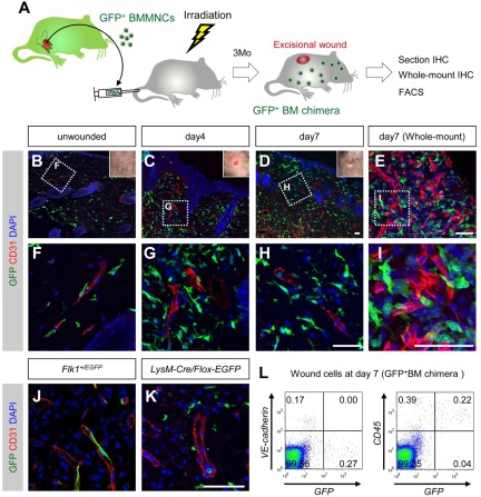
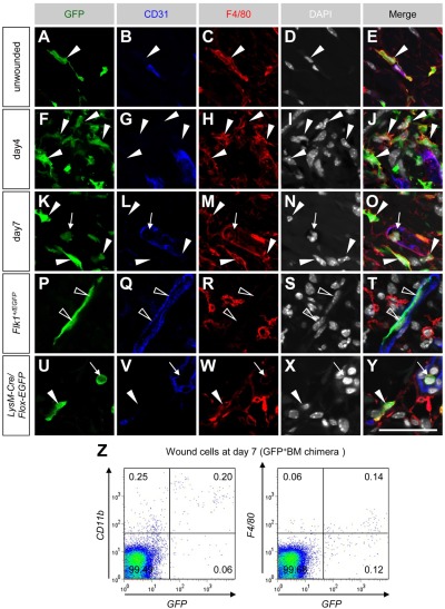
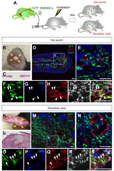
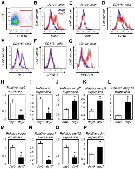
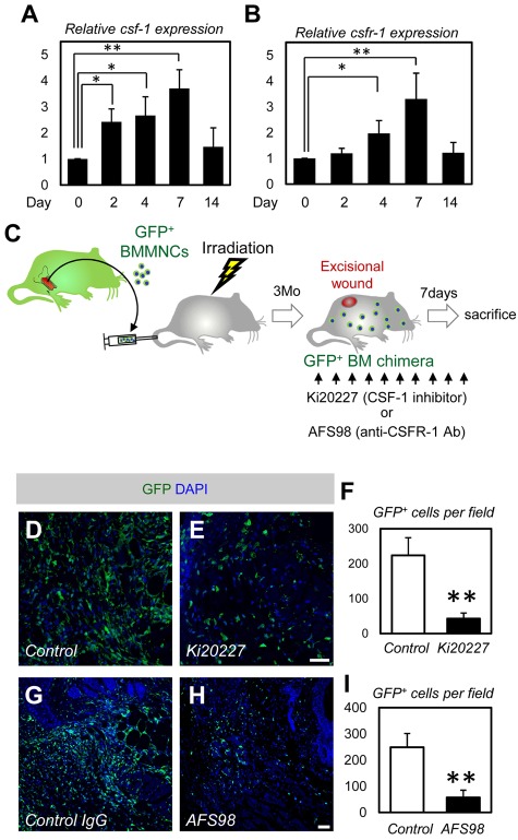
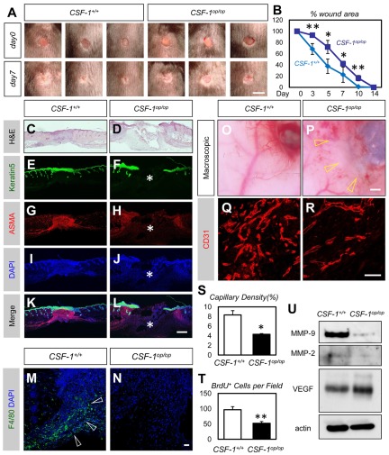
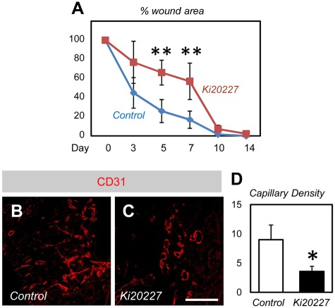
Bone marrow-derived cells (BMDCs) contribute to postnatal vascular growth by differentiating into endothelial cells or secreting angiogenic factors. However, the extent of their endothelial differentiation highly varies according to the angiogenic models used. Wound healing is an intricate process in which the skin repairs itself after injury. As a process also observed in cancer progression, neoangiogenesis into wound tissues is profoundly involved in this healing process, suggesting the contribution of BMDCs. However, the extent of the differentiation of BMDCs to endothelial cells in wound healing is unclear. In this study, using the green fluorescent protein-bone marrow chim-eric experiment and high resolution confocal microscopy at a single cell level, we observed no endothelial differentiation of BMDCs in 2 acute wound healing models (dorsal excisional wound and ear punch) and a chronic wound healing model (decubitus ulcer). Instead, a major proportion of BMDCs were macrophages. Indeed, colony-stimulating factor 1 (CSF-1) inhibition depleted approximately 80% of the BMDCs at the wound healing site. CSF-1-mutant (CSF-1(op/op)) mice showed significantly reduced neoangiogenesis into the wound site, supporting the substantial role of BMDCs as macrophages. Our data show that the proangiogenic effects of macrophages, but not the endothelial differentiation, are the major contribution of BMDCs in wound healing.


Figures

Figure 1
Figure 1
BMDCs do not differentiate into endothelial cells in dorsal excisional wounds. (A) The procedure for the dorsal excisional wound model combined with the GFP-bone marrow chimeric experiment. (B-H) Sectional immunohistochemistry for indicated antibodies. Grids in panels B through D indicate the fields in panels F through H, respectively. None of the CD31+ vascular endothelium is stained for GFP. (E,I) Whole-mount immunohistochemistry for indicated antibodies. A grid in panel E indicates the field in panel I. (J-K) Sectional immunohistochemistry for the tissues 7 days after wounding in Flk-1+/EGFP mice (J) or LysM-Cre/Flox-CAT-EGFP mice (K). GFP was detected only in endothelial cells of Flk-1+/EGFP mice and myeloid cells of LysM-Cre/Flox-CAT-EGFP mice. (L) FACS analysis for dissociated cells in wound tissues (day 7) of mice reconstituted with GFP+ bone marrow cells. None of GFP+ cells is positive for vascular endothelial-cadherin, and most GFP+ cells are positive for CD45. Bars represent 50 μm.
Figure 2
Figure 2
A major proportion of BMDCs recruited into wound tissues are macrophages in dorsal excisional wounds. (A-O) Sectional immunohistochemistry for indicated antibodies in unwounded and wounded tissues of mice reconstituted with GFP+ bone marrow cells. Most GFP+ cells located perivascularly (closed arrowheads) or in the vascular lumen (arrows) expressed F4/80. (P-Y) Sectional immunohistochemistry for the tissues 7 days after wounding in Flk-1+/EGFP mice (P-T) or LysM-Cre/Flox-CAT-EGFP mice (U-Y). GFP was detected only in endothelial cells of Flk-1+/EGFP mice (open arrowheads) and F4/80+ monocytes (arrows)/macrophages (closed arrowheads) of LysM-Cre/Flox-CAT-EGFP mice. (Z) FACS analysis for dissociated cells in wound tissues at day 7 indicated that approximately 70% of GFP+ cells are positive for CD11b, but F4/80 expression in FACS was weak and highly variable. Bar represents 50 μm.
Figure 3
Figure 3
BMDCs do not differentiate into endothelial cells in ear punch wounds and decubitus ulcers. (A) The procedure for the ear punch wound model or the decubitus ulcer model combined with the GFP-bone marrow chimeric experiment. (B-S) Macroscopic views (hematoxylin and eosin staining) or sectional immunohistochemistry for indicated antibodies. Grids in panels D and M indicate the fields in panels F-J and panels O-S, respectively. GFP+ cells that accumulated in the granulation tissues are mostly F4/80+ macrophages (arrowheads). Bars represent 50 μm.
Figure 4
Figure 4
Macrophages recruited into the wound tissues possess M2-polarized properties and express abundant MMPs. (A-H) FACS analysis for CD11b+ cells from unwounded tissues (blue lines) or dorsal excisional wounds at day 7 (red lines). Macrophages in wound tissues showed higher expression of Mrc1 (B), CD48 (D), and VEGFR1 (G) and lower expression of CD86 (C) and Tie-2 (E). (I-Q) Quantitative PCR analysis for CD11b+ cells from unwounded tissues (day 0) or dorsal excisional wounds at day 7 (n = 4). Macrophages in wound tissues showed higher expression of mmp2 (J), mmp9 (K), mmp13 (L), and csfr-1 (P) and lower expression of nos2 (H), il6 (I), vegfa (M), angpt1 (N), and cxcl-12 (O). *P < .05.
Figure 5
Figure 5
CSF-1 inhibition mostly depletes BMDCs in wound healing. (A-B) Quantitative PCR analysis for csf-1 and csfr-1 (n = 4). (C) The procedure for CSF-1 inhibition combined with the dorsal excisional wound model and the GFP-BM chimeric experiment. (D-E) Immunohistochemistry for dorsal skin wounds of PBS- or Ki20227-injected GFP-BM chimeric mice. Ki20227 treatment depleted a large number of GFP+ BMDCs in the wound site. (F) Quantification of the GFP+ cells (n = 3). (G-H) Immunohistochemistry for dorsal skin wounds of control IgGs- or AFS98-injected GFP-BM chimeric mice. AFS98 treatment depleted a large number of GFP+ BMDCs in the wound site. (I) Quantification of the GFP+ cells (n = 3). Bars represent 100 μm. *P < .05, **P < .01.
Figure 6
Figure 6
Decreased neovascularization in the wound area of CSF-1op/op mice. (A) Representative macroscopic views of full-thickness excisional wounds in the back skin of CSF-1+/+ or CSF-1op/op mice. Note delayed wound closure in CSF-1op/op mice. (B) Percentage of wound closure of dorsal excisional wounds (n = 6). (C-L) Hematoxylin and eosin staining (C-D) or immunohistochemistry of Keratin5 (green), α-smooth muscle actin (red), and 4,6-diamidino-2-phenylindole (blue) (E-L) in the healing edges at 7 days after wounding. Note delayed epidermal closure (E-F) and dermal contraction (G-H) in CSF-1op/op mice (asterisks). (M-N) Immunohistochemistry of F4/80 (green) and 4,6-diamidino-2-phenylindole (blue) in the healing edges at 7 days after wounding. Macrophages are abundantly recruited in the healing edge of CSF-1+/+ (arrowheads) but not in that of CSF-1op/op mice. (O-P) Representative macroscopic views of the ventral sides of the dorsal excisional wounds in CSF-1+/+ or CSF-1op/op mice (7 days after wounding). Note decreased vessels in the wound area of CSF-1op/op mice. (Q-R) Images for sectional immunohistochemistry of CD31. Neovascularization is decreased in the wound area of CSF-1op/op mice compared with that of CSF-1+/+ mice. (S-T) Quantification in the capillary density or the number of BrdU+ cells in the healing edges 7 days after wounding (n = 6). (U) Representative Western blotting for the wound tissues at 7 days after wounding. Although VEGF expression was not altered, MMP-9 and MMP-2 were down-regulated in CSF-1op/op mice. Bars represent 5 mm in A; 500 μm in C-L,O-P; and 50 μm in M-N,Q-R. *P < .05. **P < .01.
Figure 7
Figure 7
Delayed wound healing in Ki20227-treated mice. (A) Percentage wound closure of dorsal excisional wounds (n = 6). (B-C) Immunohistochemistry of CD31 in the healing edges at 7 days after wounding. Neovascularization is decreased in the wound area of Ki20227-treated mice compared with that of vehicle-treated mice. (D) Quantification in the capillary density in the healing edges 7 days after wounding (n = 6). Bar represents 50 μm. *P < .05. **P < .01.

References

    1. Risau W. Mechanisms of angiogenesis. Nature. 1997;386(6626):671–674. - PubMed
    1. Asahara T, Murohara T, Sullivan A, et al. Isolation of putative progenitor endothelial cells for angiogenesis. Science. 1997;275(5302):964–967. - PubMed
    1. Bertolini F, Shaked Y, Mancuso P, Kerbel RS. The multifaceted circulating endothelial cell in cancer: towards marker and target identification. Nat Rev Cancer. 2006;6(11):835–845. - PubMed
    1. Kerbel RS. Tumor angiogenesis. N Engl J Med. 2008;358(19):2039–2049. - PMC - PubMed
    1. Gao D, Nolan DJ, Mellick AS, Bambino K, McDonnell K, Mittal V. Endothelial progenitor cells control the angiogenic switch in mouse lung metastasis. Science. 2008;319(5860):195–198. - PubMed

Publication types