. 2011;194(6):521-30.
doi: 10.1159/000323875. Epub 2011 Mar 18.

Sensory innervation of the nonspecialized connective tissues in the low back of the rat

Affiliations

Sensory innervation of the nonspecialized connective tissues in the low back of the rat

Sarah M Corey et al. Cells Tissues Organs. 2011.

Abstract

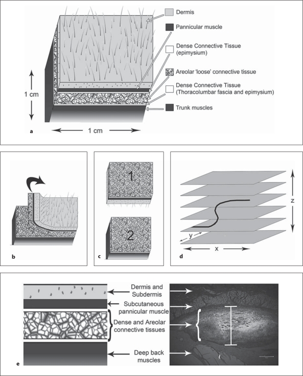
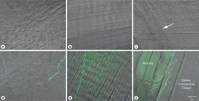
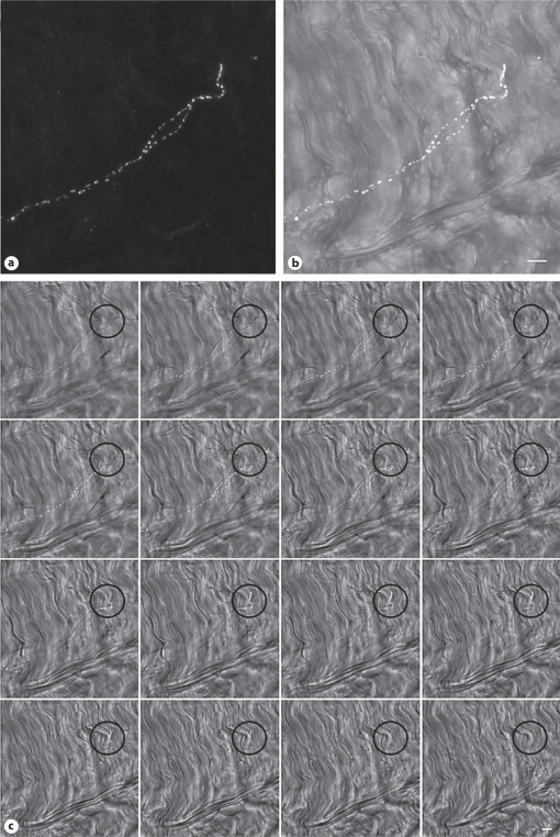
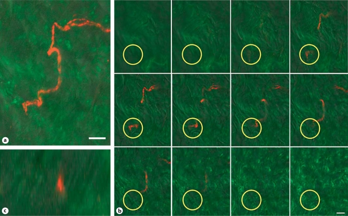
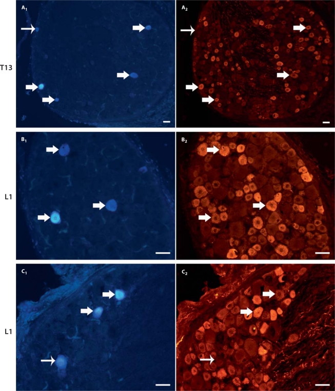
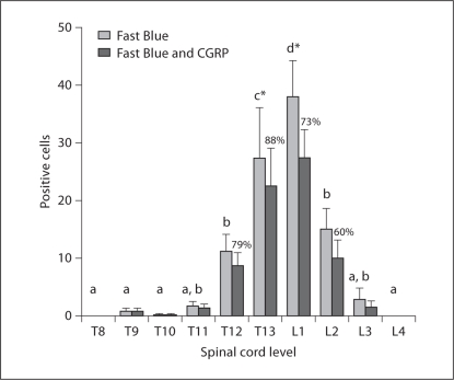
Chronic musculoskeletal pain, including low back pain, is a worldwide debilitating condition; however, the mechanisms that underlie its development remain poorly understood. Pathological neuroplastic changes in the sensory innervation of connective tissue may contribute to the development of nonspecific chronic low back pain. Progress in understanding such potentially important abnormalities is hampered by limited knowledge of connective tissue's normal sensory innervation. The goal of this study was to evaluate and quantify the sensory nerve fibers terminating within the nonspecialized connective tissues in the low back of the rat. With 3-dimensional reconstructions of thick (30-80 μm) tissue sections we have for the first time conclusively identified sensory nerve fiber terminations within the collagen matrix of connective tissue in the low back. Using dye labeling techniques with Fast Blue, presumptive dorsal root ganglia cells that innervate the low back were identified. Of the Fast Blue-labeled cells, 60-88% also expressed calcitonin gene-related peptide (CGRP) immunoreactivity. Based on the immunolabeling with CGRP and the approximate size of these nerve fibers (≤2 μm) we hypothesize that they are Aδ or C fibers and thus may play a role in the development of chronic pain.


Figures

Fig. 1
Fig. 1
Methods for identification of nerve endings. a Anatomy of the tissue layers of the low back including skin, subcutaneous muscle, dense connective tissue, areolar connective tissue, dense connective tissue, and the deep back muscles. b, c For thick tissue sectioning, the areolar and dense connective tissues were isolated by separating the tissue block as shown. d Collection of a stack of images through the z plane (orthogonal to the section) with confocal microscopy allows for nerve fibers and their surrounding tissues to be evaluated. e Histological section of tissues of the low back in the region of Fast Blue injection. Fast Blue is visualized in UV and is white in this image. The vertical bar at the center of the image indicates the spread of tracer within the areolar and dense connective tissues and not into the muscle tissue or dermis. Scale bar = 200 μm.
Fig. 2
Fig. 2
PGP9.5 (pan neuronal marker) labeling and differential interference contrast (DIC) imaging. Single slices from confocal optical z stacks of areolar connective tissue (a) dense connective tissue (b) and muscle (c) in the low back of the rat visualized with DIC. Examples of DIC combined with PGP9.5 antibody labeling of areolar connective tissue (d), dense connective tissue (e) and muscle and dense connective tissue (f). Lens: PlanApochromat ×20 (0.75 N.A.). Scale bar = 50 μm.
Fig. 3
Fig. 3
Confocal z series of 34 optical slices representing 25.4 μm of tissue. a Full projection image of the CGRP-positive nerve fiber (white). b Projection of a subset of optical tissue slices (left to right) to visualize the nerve fiber within the collagen matrix with DIC (gray scale). c Gallery of individual slices (every other slice) from the optical z stack demonstrating collagen above and below the CGRP sensory nerve fiber. Black circles indicate the location of the nerve ending. The lens was a PlanApochromat ×25 (0.8 NA) with a zoom factor of 2.6. Scale bars = 10 μm.
Fig. 4
Fig. 4
Confocal z series of 25 sections representing 35.4 μm of tissue. a Projection image of the 9th to 18th optical tissue sections (13.3 μm thickness) to show the full nerve fiber. Sensory nerve is labeled with CGRP (red) and collagen is labeled with antibody (collagen-1; green) and visualized with DIC (gray scale). b Gallery (left to right) showing every other slice from the optical z stack demonstrating collagen above and below the CGRP-expressing sensory nerve fiber. Yellow circles outline the termination of the nerve fiber. c Orthogonal view of the z plane of the tissue at the nerve ending. The lens used was a PlanApochromat ×20 (0.75 NA) with a zoom factor of 2.6. Scale bars = 10 μm.
Fig. 5
Fig. 5
Fast Blue retrograde tracer and CGRP expression within the DRG. Examples of DRG tissue sections labeled with Fast Blue (A1, B1, and C1) and corresponding staining of each section with CGRP (A2, B2, and C2). Thick arrows indicate cells labeled with both Fast Blue and CGRP and thin arrows demonstrate Fast Blue-positive cell bodies that do not express CGRP. DRG levels are T13 for section A and L1 for sections B and C. Scale bars = 50 μm.
Fig. 6
Fig. 6
Quantitative analysis of Fast Blue- and CGRP-labeled cells at T8-L4 spinal cord levels. Light gray bars indicate the distribution of counted Fast Blue-labeled cells (mean with SEM) innervating the connective tissues (tracer injected lateral to L3). Columns sharing a letter are not significantly different from each other. Spinal cord levels indicated by (∗) are significantly different from all other levels (Fisher's LSD; p < 0.05). The expression of CGRP (dark gray bars) within the population of Fast Blue-labeled cells (mean with SEM) is indicated by the percentages over the dark gray bars.

References

    1. Bednar D.A., Orr F.W., S. G.T. Observations on the pathomorphology of the thoracolumbar fascia in chronic mechanical back pain: a microscopic study. Spine. 1995;20:1161–1164. - PubMed
    1. Bjur D., Alfredson H., Forsgren S. The innervation pattern of the human Achilles tendon: studies of the normal and tendinosis tendon with markers for general and sensory innervation. Cell Tissue Res. 2005;320:201–206. - PubMed
    1. Borg-Stein J., Wilkins A. Soft tissue determinants of low back pain. Curr Pain Headache Rep. 2006;10:339–344. - PubMed
    1. Bove G.M., Light A.R. Unmyelinated nociceptors of rat paraspinal tissues. J Neurophysiol. 1995;73:1752–1762. - PubMed
    1. Box G., Hunter W., Hunter J. Statistics for Experimenters. New York: Wiley; 1978.

Publication types

Substances