. 2011 Apr;70(4):302-13.
doi: 10.1097/NEN.0b013e31821350b0.

Dysferlin, annexin A1, and mitsugumin 53 are upregulated in muscular dystrophy and localize to longitudinal tubules of the T-system with stretch

Affiliations

Dysferlin, annexin A1, and mitsugumin 53 are upregulated in muscular dystrophy and localize to longitudinal tubules of the T-system with stretch

Leigh B Waddell et al. J Neuropathol Exp Neurol. 2011 Apr.

Abstract

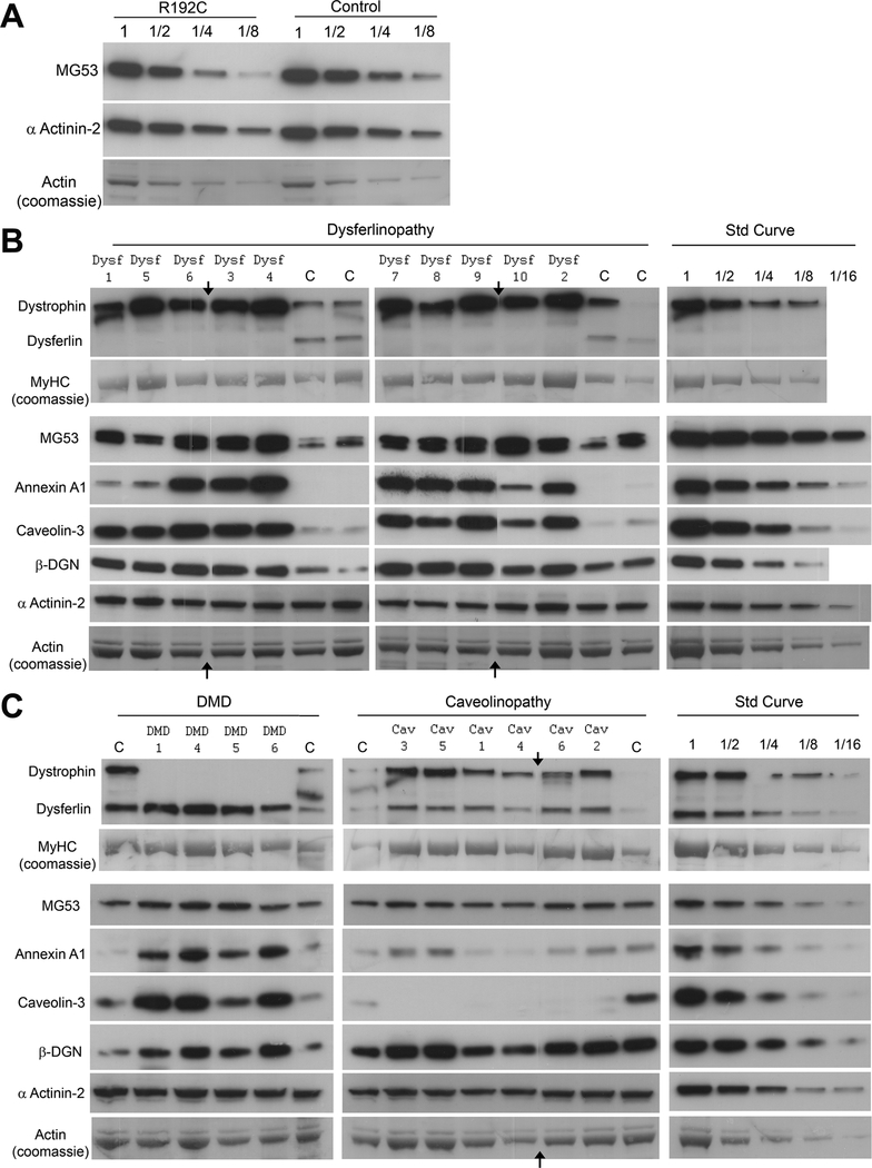
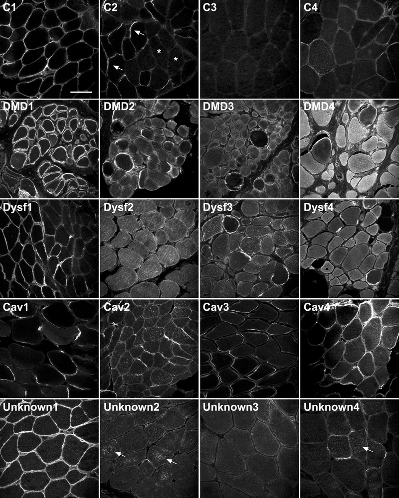
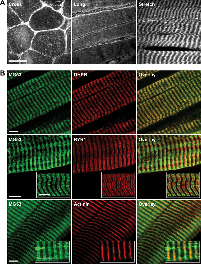
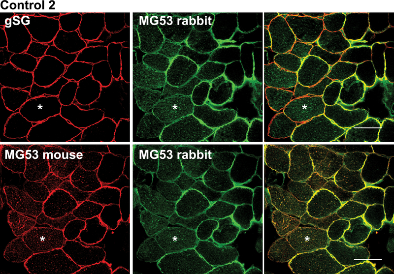
Mutations in dysferlin cause an inherited muscular dystrophy because of defective membrane repair. Three interacting partners of dysferlin are also implicated in membrane resealing: caveolin-3 (in limb girdle muscular dystrophy type 1C), annexin A1, and the newly identified protein mitsugumin 53 (MG53). Mitsugumin 53 accumulates at sites of membrane damage, and MG53-knockout mice display a progressive muscular dystrophy. This study explored the expression and localization of MG53 in human skeletal muscle, how membrane repair proteins are modulated in various forms of muscular dystrophy, and whether MG53 is a primary cause of human muscle disease. Mitsugumin 53 showed variable sarcolemmal and/or cytoplasmic immunolabeling in control human muscle and elevated levels in dystrophic patients. No pathogenic MG53 mutations were identified in 50 muscular dystrophy patients, suggesting that MG53 is unlikely to be a common cause of muscular dystrophy in Australia. Western blot analysis confirmed upregulation of MG53, as well as of dysferlin, annexin A1, and caveolin-3 to different degrees, in different muscular dystrophies. Importantly, MG53, annexin A1, and dysferlin localize to the t-tubule network and show enriched labeling at longitudinal tubules of the t-system in overstretch. Our results suggest that longitudinal tubules of the t-system may represent sites of physiological membrane damage targeted by this membrane repair complex.


Figures

Figure 1.
Figure 1.
Mitsugumin-53 (MG53) has variable sarcolemmal and/or cytoplasmic immunolabeling in control human muscle samples and increased staining in dystrophic patient muscle samples. Immunohistochemical analysis of MG53 (rabbit polyclonal MG53 antibody) in 4 human control skeletal muscle samples (C1–C4), 4 Duchenne muscular dystrophy human skeletal muscle samples (DMD1-DMD4), 4 Dysferlin human skeletal muscle samples (DYSF1-DYSF4), 4 Caveolin-3 human skeletal muscle samples (CAV1-CAV4), and 4 unknown limb girdle muscular dystrophy human skeletal muscle samples (Unknown1-Unknown4). Arrows in C2 indicate fibers with enriched membrane staining for MG53; stars in C2 indicate fibers within the same section showing predominantly cytoplasmic MG53 labelling. Arrows in Unknown 2 and Unknown 4 indicate punctate cytoplasmic labelling of MG53 aggregates. All samples were stained on the same day, under the same conditions. All imaging was completed over 2 days using identical confocal settings. Scale bar = 100 μm.
Figure 2:
Figure 2:
Two distinct anti-mitsugumin-53 (MG53) antibodies confirm variable localization of MG53 in control human muscle. Sequential muscle cryosections from Control 2 were labelled with either rabbit polyclonal anti-MG53 plus a mouse-monoclonal recognizing γ-sarcoglycan (to demonstrate membrane fidelity in fibers that did not show MG53 sarcolemmal labelling) (top row), or mouse monoclonal anti-MG53 plus rabbit polyclonal anti-MG53 antibodies (bottom row). Both antibodies show a highly similar staining pattern for MG53, which variably localizes to sarcolemma or cytoplasm (asterisks) in different fibers. Bar = 50 μm.
Figure 3.
Figure 3.
Membrane repair proteins are upregulated in muscular dystrophy. Western blot analysis of muscle biopsies from patients with muscular dystrophy and age-matched controls. (A) Serial dilutions of total protein lysates from muscle of an undiagnosed limb girdle muscular dystrophy (LGMD) patient who is heterozygous for the mitsugumin-53 (MG53) p.R192C polymorphism, and an age-matched control, demonstrating equivalent levels of MG53 protein. (B) Ten LGMD2B (dysferlinopathy) and 4 age-matched control samples. (C) Four Duchenne muscular dystrophy (DMD) and 6 LGMD1C (caveolinopathy) samples with age-matched controls. Arrows on B and C indicate where a lane with a degraded sample was removed for image presentation. Standard curves for each protein were generated from serial 2-fold dilutions of a control sample (Std Curve). Labels: Dysf 1, DMD 1, Cav 1 etc. correspond to samples in Figure 1; MyHC = myosin heavy chain; β-DGN = β-dystroglycan.
Figure 4.
Figure 4.
Mitsugumin-53 (MG53) immunolocalizes to t-tubules in stretched human skeletal muscle. (A) Immunohistochemical analysis of MG53 in human control (C1) skeletal muscle cut in cross-section (Cross) in longitudinal section (Long) and in stretched longitudinal section (Stretch). A striated pattern is most evident in stretched longitudinal muscle. Bar = 50 μm. (B) Immunohistochemical analysis of MG53, dihydropyridine receptor (DHPR), dihydropyridine receptor (RYR1) and α-actinin in stretched longitudinal human control muscle (C1) to investigate the localization of MG53. The MG53 striated staining pattern overlaps with DHPR staining, but not with RYR1 or actinin staining. Bars = 10 μm.
Figure 5.
Figure 5.
Membrane repair proteins label t-tubule and longitudinal t-tubules in control stretched muscle. Immunohistochemical analysis of mitsugumin-53 (MG53) with spectrin, dysferlin, annexin-A1 and caveolin-3 in longitudinal human control (C1) skeletal muscle stretched at room temperature before freezing. Arrows indicate enriched co-labelling of MG53 and dysferlin at the junction of longitudinal tubules with the t-system. Stars indicate co-labelling of MG53 and annexin A1 at longitudinal tubules of the t-system. Scale bars = 10 μm.

References

    1. Nowak KJ, Davies KE. Duchenne muscular dystrophy and dystrophin: pathogenesis and opportunities for treatment. EMBO Rep 2004:5;872–6 - PMC - PubMed
    1. Bansal D, Miyake K, Vogel SS, et al. Defective membrane repair in dysferlin-deficient muscular dystrophy. Nature 2003:423;168–72 - PubMed
    1. Terasaki M, Miyake K, McNeil PL. Large plasma membrane disruptions are rapidly resealed by Ca2+-dependent vesicle-vesicle fusion events. J Cell Biol 1997:139;63–74 - PMC - PubMed
    1. Matsuda C, Hayashi YK, Ogawa M, et al. The sarcolemmal proteins dysferlin and caveolin-3 interact in skeletal muscle. Hum Mol Genet 2001:10;1761–6 - PubMed
    1. Woodman SE, Sotgia F, Galbiati F, et al. Caveolinopathies: mutations in caveolin-3 cause four distinct autosomal dominant muscle diseases. Neurology 2004:62;538–43 - PubMed

Publication types

MeSH terms