. 2011 Jul;18(7):637-45.
doi: 10.1038/gt.2011.3. Epub 2011 Mar 17.

AAV-mediated photoreceptor transduction of the pig cone-enriched retina

Affiliations
Free PMC article

AAV-mediated photoreceptor transduction of the pig cone-enriched retina

C Mussolino et al. Gene Ther. 2011 Jul.
Free PMC article

Abstract

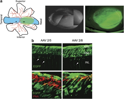
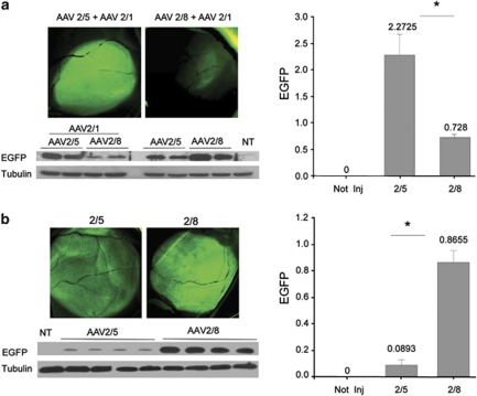
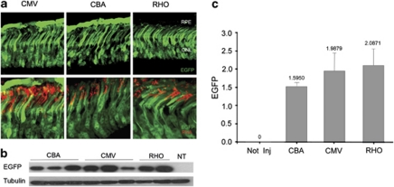
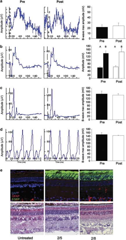
Recent success in clinical trials supports the use of adeno-associated viral (AAV) vectors for gene therapy of retinal diseases caused by defects in the retinal pigment epithelium (RPE). In contrast, evidence of the efficacy of AAV-mediated gene transfer to retinal photoreceptors, the major site of inherited retinal diseases, is less robust. In addition, although AAV-mediated RPE transduction appears efficient, independently of the serotype used and species treated, AAV-mediated photoreceptor gene transfer has not been systematically investigated thus so far in large animal models, which also may allow identifying relevant species-specific differences in AAV-mediated retinal transduction. In the present study, we used the porcine retina, which has a high cone/rod ratio. This feature allows to properly evaluate both cone and rod photoreceptors transduction and compare the transduction characteristics of AAV2/5 and 2/8, the two most efficient AAV vector serotypes for photoreceptor targeting. Here we show that AAV2/5 and 2/8 transduces both RPE and photoreceptors. AAV2/8 infects and transduces photoreceptor more efficiently than AAV2/5, similarly to what we have observed in the murine retina. The use of the photoreceptor-specific rhodopsin promoter restricts transgene expression to porcine rods and cones, and results in photoreceptor transduction levels similar to those obtained with the ubiquitous promoters tested. Finally, immunological, toxicological and biodistribution studies support the safety of AAV subretinal administration to the large porcine retina. The data presented here on AAV-mediated transduction of the cone-enriched porcine retina may affect the development of gene-based therapies for rare and common severe photoreceptor diseases.


Figures

Figure 1
Figure 1
Tropism of AAV2/5 and AAV2/8 vectors subretinally injected in the porcine retina. (a, right panel) Schematic representation of the visual streak in the porcine retina with highest cone density (light blue; re-drawing from reference 24). The light green area represents the site and the extension of subretinal injection. (a, middle panel) Assembly of black and white fundus images showing dome-like retinal bleb following subretinal injection of 100 μl of AAV vector containing- solution. The asterisk indicates the injection site. (b) Confocal fluorescence microscopy analysis of retinal sections at 6 weeks after subretinal injection of AAV2/5-, or AAV2/8-CMV-EGFP. (b, upper panel) Outer retinal EGFP expression appearance ( × 20 magnification). Arrows indicate EGFP-positive cells in the inner nuclear layer (INL). The images of the INL were taken at higher exposure than those of the outer retina. RPE, retinal pigment epithelium; ONL, outer nuclear layer (scale bar=10 μm). (b, lower panels) Cone transduction by the different AAV serotypes. Co-localization of EGFP expression and PNA-lectin staining (cone sheats in red) evaluated by confocal microscopy (scale bar=100 μm).
Figure 2
Figure 2
AAV-mediated transduction of the porcine retina. (a) Fundus fluorescence imaging of the whole-mounted retinas showed higher EGFP expression in AAV2/5- than AAV2/8 treated retinas when co-injected with AAV2/1 (Figure 1a, upper panels). (a, Lower panel) Western blot analysis with anti-EGFP (upper blot) or anti-β-tubulin (lower blot) antibodies of lysates from swine retinas transduced with AAV-2/5, -2/8 (n=4 for each serotype). (a, Lower right) Histogram representing the EGFP expression levels measured by western blot. In the histogram values of EGFP (pixels inches−1) normalized against β-tubulin (pixels inches−1) are reported (n=4 eyes per group; *AAV2/5 vs AAV2/8: P=0.0098). (b) Fundus appearance (upper left) and quantification of the EFGP expression levels (upper right and lower panels) by western blot analysis of porcine retinas injected with AAV2/5-CMV-EGFP or AAV2/8-CMV-EGFP alone (n=4 eyes per group; *AAV2/5 vs AAV2/8: P=0.00014).
Figure 3
Figure 3
Subretinal injections of AAV2/5 vectors expressing EGFP from ubiquitous and photoreceptor-specific promoters. (a, Upper panels) Confocal fluorescence microscopy images ( × 63 magnification) of retinal sections showing EGFP expression from cytomegalovirus (CMV), Chicken-β-actin (CBA) or human rhodopsin (RHO) promoters (Scale bar=100 μm). (a, Lower panels) Cone transduction is indicated by the co-localization of EGFP expression and PNA staining (arrows). RPE, retinal pigment epithelium; ONL, outer nuclear layer (scale bar=100 μm). (b) Western blot analysis with anti-EGFP (upper blot) or anti-β-tubulin (lower blot) antibodies of lysates from porcine retinas transduced with AAV-2/5-EGFP driven by CMV, CBA or RHO promoters. (c) In the left histogram, values of EGFP (pixels inches−1) normalized against β-tubulin (pixels inches−1) are reported (n=3 for each serotype).
Figure 4
Figure 4
Safety of AAV vector delivery to porcine retinas. Electroretinographic recordings at baseline (pretreatment) and at killing (post treatment). (a–d) Comparison of the tracings derived from representative eyes before and after subretinal injections of AAV vectors. The histograms show the maximum B-wave average response of all recorded eyes (n=6 eyes pre-treatment; n=5 eyes post treatment). (a) Rod responses (dark-adapted conditions); (b) Maximal response (A-wave and B-wave) of rods and cones (dark-adapted conditions, single white flash); (c) Cone responses isolated using a bright single flash (light-adapted conditions, background white light); (d) Cone responses isolated using a flicker response (light-adapted conditions). (e) Immunofluorescence image ( × 20 magnification) of retinal sections from animals injected with AAV2/5 or 2/8 and stained with glial fibrillary acidic protein (GFAP; in red) as marker of gliosis. EGFP expression (in green) shows the transduced area. RPE, retinal pigment epithelium; ONL, outer nuclear layer; INL, inner nuclear layer (scale bar=10 μm).

References

    1. Cremers FP, van den Hurk JA, den Hollander AI. Molecular genetics of Leber congenital amaurosis. Hum Mol Genet. 2002;11:1169–1176. - PubMed
    1. Bainbridge JW, Smith AJ, Barker SS, Robbie S, Henderson R, Balaggan K, et al. Effect of gene therapy on visual function in Leber's congenital amaurosis. N Engl J Med. 2008;358:2231–2239. - PubMed
    1. Cideciyan AV, Aleman TS, Boye SL, Schwartz SB, Kaushal S, Roman AJ, et al. Human gene therapy for RPE65 isomerase deficiency activates the retinoid cycle of vision but with slow rod kinetics. Proc Natl Acad Sci USA. 2008;105:15112–15117. - PMC - PubMed
    1. Cideciyan AV, Hauswirth WW, Aleman TS, Kaushal S, Schwartz SB, Boye SL, et al. Human RPE65 gene therapy for Leber congenital amaurosis: persistence of early visual improvements and safety at 1 year. Hum Gene Ther. 2009;20:999–1004. - PMC - PubMed
    1. Cideciyan AV, Hauswirth WW, Aleman TS, Kaushal S, Schwartz SB, Boye SL, et al. Vision 1 year after gene therapy for Leber's congenital amaurosis. N Engl J Med. 2009;361:725–727. - PMC - PubMed

Publication types

Supplementary concepts