

TDP-43 regulates Drosophila neuromuscular junctions growth by modulating Futsch/MAP1B levels and synaptic microtubules organization
- PMID: 21412434
- PMCID: PMC3055892
- DOI: 10.1371/journal.pone.0017808
TDP-43 regulates Drosophila neuromuscular junctions growth by modulating Futsch/MAP1B levels and synaptic microtubules organization
Abstract
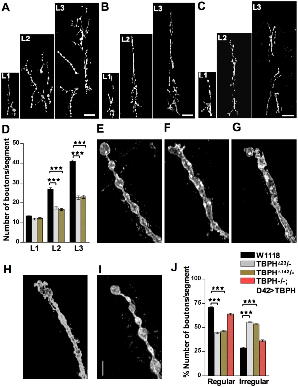
TDP-43 is an evolutionarily conserved RNA binding protein recently associated with the pathogenesis of different neurological diseases. At the moment, neither its physiological role in vivo nor the mechanisms that may lead to neurodegeneration are well known. Previously, we have shown that TDP-43 mutant flies presented locomotive alterations and structural defects at the neuromuscular junctions. We have now investigated the functional mechanism leading to these phenotypes by screening several factors known to be important for synaptic growth or bouton formation. As a result we found that alterations in the organization of synaptic microtubules correlate with reduced protein levels in the microtubule associated protein futsch/MAP1B. Moreover, we observed that TDP-43 physically interacts with futsch mRNA and that its RNA binding capacity is required to prevent futsch down regulation and synaptic defects.
Conflict of interest statement
Figures


Similar articles
-
A presynaptic role of microtubule-associated protein 1/Futsch in Drosophila: regulation of active zone number and neurotransmitter release.J Neurosci. 2014 May 14;34(20):6759-71. doi: 10.1523/JNEUROSCI.4282-13.2014. J Neurosci. 2014. PMID: 24828631 Free PMC article.
-
TBPH/TDP-43 modulates translation of Drosophila futsch mRNA through an UG-rich sequence within its 5'UTR.Brain Res. 2016 Sep 15;1647:50-56. doi: 10.1016/j.brainres.2016.02.022. Epub 2016 Feb 18. Brain Res. 2016. PMID: 26902497
-
Futsch/MAP1B mRNA is a translational target of TDP-43 and is neuroprotective in a Drosophila model of amyotrophic lateral sclerosis.J Neurosci. 2014 Nov 26;34(48):15962-74. doi: 10.1523/JNEUROSCI.2526-14.2014. J Neurosci. 2014. PMID: 25429138 Free PMC article.
-
The Drosophila microtubule associated protein Futsch is phosphorylated by Shaggy/Zeste-white 3 at an homologous GSK3beta phosphorylation site in MAP1B.Mol Cell Neurosci. 2006 Oct;33(2):188-99. doi: 10.1016/j.mcn.2006.07.004. Epub 2006 Sep 1. Mol Cell Neurosci. 2006. PMID: 16949836
-
Drosophila Futsch regulates synaptic microtubule organization and is necessary for synaptic growth.Neuron. 2000 May;26(2):371-82. doi: 10.1016/s0896-6273(00)81170-8. Neuron. 2000. PMID: 10839356
Cited by
-
Drosophila Answers to TDP-43 Proteinopathies.J Amino Acids. 2012;2012:356081. doi: 10.1155/2012/356081. Epub 2012 Apr 18. J Amino Acids. 2012. PMID: 22577517 Free PMC article.
-
The microtubule cytoskeleton at the synapse.Neurosci Lett. 2021 May 14;753:135850. doi: 10.1016/j.neulet.2021.135850. Epub 2021 Mar 26. Neurosci Lett. 2021. PMID: 33775740 Free PMC article. Review.
-
TDP-43 proteinopathy and mitochondrial abnormalities in neurodegeneration.Mol Cell Neurosci. 2019 Oct;100:103396. doi: 10.1016/j.mcn.2019.103396. Epub 2019 Aug 21. Mol Cell Neurosci. 2019. PMID: 31445085 Free PMC article. Review.
-
Neurodegeneration the RNA way.Prog Neurobiol. 2012 May;97(2):173-89. doi: 10.1016/j.pneurobio.2011.10.006. Epub 2011 Nov 3. Prog Neurobiol. 2012. PMID: 22079416 Free PMC article. Review.
-
Neurotoxicity of the Cyanotoxin BMAA Through Axonal Degeneration and Intercellular Spreading.Neurotox Res. 2018 Jan;33(1):62-75. doi: 10.1007/s12640-017-9790-1. Epub 2017 Aug 25. Neurotox Res. 2018. PMID: 28842862
References
-
- Buratti E, Baralle FE. The molecular links between TDP-43 dysfunction and neurodegeneration. Adv Genet. 2009;66:1–34. - PubMed
-
- Strong MJ. The evidence for altered RNA metabolism in amyotrophic lateral sclerosis (ALS). J Neurol Sci. 288:1–12. - PubMed
-
- Buratti E, Baralle FE. Characterization and functional implications of the RNA binding properties of nuclear factor TDP-43, a novel splicing regulator of CFTR exon 9. J Biol Chem. 2001;276:36337–36343. - PubMed
-
- Moisse K, Volkening K, Leystra-Lantz C, Welch I, Hill T, et al. Divergent patterns of cytosolic TDP-43 and neuronal progranulin expression following axotomy: implications for TDP-43 in the physiological response to neuronal injury. Brain Res. 2009;1249:202–211. - PubMed
-
- Neumann M, Sampathu DM, Kwong LK, Truax AC, Micsenyi MC, et al. Ubiquitinated TDP-43 in frontotemporal lobar degeneration and amyotrophic lateral sclerosis. Science. 2006;314:130–133. - PubMed
Publication types
MeSH terms
Substances
LinkOut - more resources
Full Text Sources
Molecular Biology Databases
Research Materials
