. 2011 Mar 11;6(3):e17748.
doi: 10.1371/journal.pone.0017748.

A live-attenuated HSV-2 ICP0 virus elicits 10 to 100 times greater protection against genital herpes than a glycoprotein D subunit vaccine

Affiliations

A live-attenuated HSV-2 ICP0 virus elicits 10 to 100 times greater protection against genital herpes than a glycoprotein D subunit vaccine

William P Halford et al. PLoS One. .

Abstract

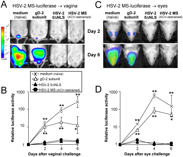
Glycoprotein D (gD-2) is the entry receptor of herpes simplex virus 2 (HSV-2), and is the immunogen in the pharmaceutical industry's lead HSV-2 vaccine candidate. Efforts to prevent genital herpes using gD-2 subunit vaccines have been ongoing for 20 years at a cost in excess of $100 million. To date, gD-2 vaccines have yielded equivocal protection in clinical trials. Therefore, using a small animal model, we sought to determine if a live-attenuated HSV-2 ICP0⁻ virus would elicit better protection against genital herpes than a gD-2 subunit vaccine. Mice immunized with gD-2 and a potent adjuvant (alum+monophosphoryl lipid A) produced high titers of gD-2 antibody. While gD-2-immunized mice possessed significant resistance to HSV-2, only 3 of 45 gD-2-immunized mice survived an overwhelming challenge of the vagina or eyes with wild-type HSV-2 (MS strain). In contrast, 114 of 115 mice immunized with a live HSV-2 ICP0⁻ virus, 0ΔNLS, survived the same HSV-2 MS challenges. Likewise, 0ΔNLS-immunized mice shed an average 125-fold less HSV-2 MS challenge virus per vagina relative to gD-2-immunized mice. In vivo imaging demonstrated that a luciferase-expressing HSV-2 challenge virus failed to establish a detectable infection in 0ΔNLS-immunized mice, whereas the same virus readily infected naïve and gD-2-immunized mice. Collectively, these results suggest that a HSV-2 vaccine might be more likely to prevent genital herpes if it contained a live-attenuated HSV-2 virus rather than a single HSV-2 protein.


Conflict of interest statement

Competing Interests: The authors have read the journal's policy and have the following conflicts: William Halford is a co-author on United States Patent Appication Publication US2010/0226940 A1, which describes the uses of herpes simplex virus mutant ICP0 in the design of a live-attenuated HSV-2 vaccine strain. This does not alter the authors' adherence to all PLoS ONE policies on sharing data and materials.

Figures

Figure 1
Figure 1. HSV-2 0ΔNLS is avirulent and immunogenic in female ICR mice.
Duration of survival following inoculation of naïve mice with culture medium containing 25,000 pfu per µl of (A) HSV-2 MS or (B) HSV-2 0ΔNLS following placement of 4 µl on left and right scarified eyes; 5 µl in left and right nostrils; 50 µl in left and right, rear footpads; or 20 µl instilled into the vaginal vault (n = 10 mice per group). A single asterisk (*) denotes a probability, p, <0.05 and a double asterisk (**) denotes p<0.001 that matched pairs of mice inoculated with (A) HSV-2 MS or (B) HSV-2 0ΔNLS survived at equivalent frequencies, as calculated by Fisher's Exact Test. (C) Mean ± sem abundance of gD-2 specific IgG antibody in mouse serum on Day 50 p.i., as determined by ELISA on 1∶100 dilutions of mouse serum (n = 10 per 0ΔNLS-immunization group; n = 5 MS-immunized mice). The y-axis represents relative units of IgG abundance expressed as “fold-increase above background,” as determined relative to a 0.33-log dilution series of high titer anti-HSV-2 antiserum that provided the standard curve that defined the quantitative relationship between anti-gD-2 IgG antibody abundance and the colorimetric development in each well of the ELISA plate (i.e., the standard curve had a goodness-of-fit of r2 = 0.99). A double asterisk (**) denotes a probability, p, <0.001 that gD-2-antibody levels were equivalent to naïve mice, as determined by one-way ANOVA and Tukey's post hoc t-test.
Figure 2
Figure 2. Mice immunized with HSV-2 0ΔNLS are resistant to HSV-2 vaginal challenge.
Mice were treated with 2 mg medoxyprogesterone 7 and 3 days prior to vaginal HSV-2 challenge . On Day 56 p.i., HSV-2 0ΔNLS- and MS-immunized mice were challenged with 500,000 pfu per vagina of HSV-2 MS. (A) HSV-2 shedding from the vagina between Days 2 and 6 post-challenge in naïve mice (n = 10) versus mice inoculated in the rear footpads with HSV-2 MS (n = 5) or HSV-2 0ΔNLS (n = 5). (B) HSV-2 shedding from the vagina of naïve mice versus mice inoculated in the eyes, nose, or vagina with HSV-2 0ΔNLS (n = 5 per group). In panels A and B, a single asterisk (*) denotes a probability, p, <0.05 and a double asterisk (**) denotes p<0.001 that HSV-2 shedding was equivalent to naïve controls on that day, as determined by one-way ANOVA and Tukey's post hoc t-test. (C) Survival frequency of naïve mice (n = 10) versus immunized mice (n = 5 per group) after HSV-2 challenge of the vagina. A double asterisk (**) denotes p<0.001 that survival frequency was equivalent to naïve mice.
Figure 3
Figure 3. Immunization with HSV-2 0ΔNLS, gD-2, or control immunogens.
(A) Design of vaccine-challenge experiments. Protein-immunized mice were injected in their right, rear footpads on Day 0 with 10 µg monophosphoryl lipid A, 2.5 µg gD-2 or GFP, and alum (n = 40 per group). On Day 30, mice received an equivalent immunization in their left, rear footpads. Virus-immunized mice received injections on Days 0 and 30 of culture medium (mock), 1×10⁶ pfu of HSV-2 0ΔNLS, or 1×10⁶ pfu of HSV-2 MS (n = 40 per group). Mice immunized with HSV-2 MS received 1 mg/ml acyclovir in drinking water from Days −1 to +20 p.i. On Day 60, blood was harvested from all mice, and on Days 80, 90, or 100, mice were challenged with wild-type HSV-2 MS. (B) HSV-2 replication in mouse footpads. In a parallel experiment, mice were footpad-injected with 1×10⁶ pfu of HSV-2 MS in the presence or absence of oral acyclovir (ACV) or 1×10⁶ pfu of HSV-2 0ΔNLS. On Days 1, 2, and 3 p.i., footpad titers of infectious HSV-2 were determined in n = 8 mice per group; on days 5 and 7 p.i., footpad titers were determined in n = 4 mice per group. All datum points represent mean ± sem pfu per footpad. A double asterisk (**) denotes p<0.001 that viral titers per footpad were the same as HSV-2 MS-inoculated mice not treated with acyclovir. (C) Mean ± sem relative abundance of gD-2 specific IgG antibody in mouse serum on Day 60 p.i., as determined by ELISA on 1∶100 dilutions of mouse serum (n = 30 per group). Relative units of IgG abundance are expressed as “fold-increase above background,” as determined relative to a 0.33-log dilution series of high titer anti-HSV-2 antiserum that provided the standard curve that defined the quantitative relationship between anti-gD-2 IgG antibody abundance and colorimetric development. A double asterisk (**) denotes p<0.001 that gD-2-antibody levels were equivalent to naïve (medium-treated) mice, as determined by one-way ANOVA and Tukey's post hoc t-test.
Figure 4
Figure 4. Resistance of naïve versus immunized mice to vaginal HSV-2 infection.
Mice were treated with 2 mg medoxyprogesterone 7 and 3 days prior to vaginal HSV-2 challenge . On Days 80, 90, or 100 p.i., mice were challenged with 500,000 pfu per vagina of HSV-2 MS (n = 5 per group). The summated results from all three experiments are presented in each panel (∑n = 15 per group). (A) Vaginal HSV-2 shedding between Days 1 and 7 post-challenge in mice that were naïve or immunized with gD-21-306t versus HSV-2 0ΔNLS. (B) Vaginal HSV-2 shedding in mice that were naïve or immunized with GFP versus HSV-2 MS. In panels A and B, a single asterisk (*) denotes p<0.05 and a double asterisk (**) denotes p<0.001 that HSV-2 shedding was equivalent to naïve mice on that day, as determined by one-way ANOVA and Tukey's post hoc t-test. (C) Mean ± sem reduction in HSV-2 shedding on Days 1–7 post-challenge relative to the average titer of HSV-2 shed by naïve mice on that day (n = 75 per group). In panel C, a single asterisk (*) denotes p<0.05 and a double asterisk (**) denotes p<0.001 that reductions in vaginal shedding of HSV-2 MS were significantly greater than a value of 1, as determined by one-way ANOVA and Tukey's post hoc t-test. The difference in reductions in HSV-2 MS vaginal shedding between 0ΔNLS- and gD-2-immunized mice was significant (p<10−23; two-sided, paired t-test). (D) Survival frequency over time following HSV-2 MS challenge of the vagina. A double asterisk (**) denotes p<0.001 that survival frequency was equivalent to naïve mice, as determined by Fisher's Exact Test. The survival rate of gD-2 immunized mice was not significantly different than naïve mice (p = 0.22, Fisher's Exact Test).
Figure 5
Figure 5. Polyclonal HSV-2 IgG antibody response elicited by HSV-2 0ΔNLS, gD-2, or control immunogens.
(A) Mean ± sem neutralizing antibody titer of Day 60 serum samples (n = 20 per group). The titer of each serum sample was considered to be the reciprocal of the largest serum dilution that reduced HSV-2's cytopathic effect in Vero cell monolayers by at least 50%. (B) Representative immunofluorescent labeling of fixed HSV-2 plaques with a 1∶5,000 dilution of Day 60 serum from each immunization group. (C) Flow cytometric measurement of pan-HSV-2-specific IgG levels in Day 60 sera, as determined by IgG binding to fixed HSV-2-infected cells versus uninfected Vero cells (n = 8 per group). In panels A and C, a double asterisk (**) denotes p<0.001 that neutralizing antibody titers or pan-HSV-2 IgG levels were equivalent to naïve mice, as determined by one-way ANOVA and Tukey's post hoc t-test. The difference in pan HSV-2 IgG levels between 0ΔNLS- and gD-2-immunized mice was significant (p<0.0001; two-sided, paired t-test). (D) Regression analysis of the logarithm of pan-HSV-2 IgG levels (x-variable, as measured on Day 60) in n = 25 mice versus the logarithmic reduction in vaginal HSV-2 MS shedding (y-variable, as measured on Day 81) observed in the same n = 25 mice at 24 hours post-vaginal challenge. The x-variable data is based on a subset of the data summarized in Figure 5C, and likewise the y-variable data is based on a subset of the data summarized in Figure 4C. The individual datum points are derived from n = 5 mice per group that were immunized with medium (naïve), GFP, gD-2, HSV-2 0ΔNLS, or HSV-2 MS (ACV-restrained infection), as indicated in the legend in Panel D. The quantity on the y-axis, Δlog (pfu/vagina), represents the logarithmic decrease of HSV-2 MS shed from an individual mouse vagina at 24 hours post-challenge relative to 5.20 logs, which was the average titer of HSV-2 MS shed by naïve mice at 24 hours post-challenge. The goodness-of-fit of the correlation between log (pan-HSV-2 IgG) and Δlog (pfu/vagina) was r2 = 0.83 and the slope of the correlation was 1.38±0.13 (p<10−9).
Figure 6
Figure 6. Vaccine-induced protection against HSV-2 MS-luciferase infection.
(A and C) Mice were treated with 2 mg medoxyprogesterone 7 and 3 days prior to vaginal HSV-2 challenge . On Day 130 p.i., mice were challenged with (A) 500,000 pfu per vagina or (C) 100,000 pfu per eye of HSV-2 MS-luciferase, and were anaesthetized and injected with 3 mg D-luciferin substrate at times post-challenge for imaging in a bioluminescent imager. Not shown in panels A or C are the age- and sex-matched, uninfected control mice included in these analyses that were anaesthetized and injected with 3 mg D-luciferin substrate at the same time, and which served as a background control to define the background level of light emission recorded from each mouse by the bioluminescent imager. (B and D) Mean ± sem of luciferase activity in mice challenged in the (B) vagina or (D) eyes with HSV-2 MS-luciferase, as measured by the fold-increase in light emission from each mouse relative to an uninfected background control mouse injected with 3 mg D-luciferin substrate. In the vaginally challenged group, each datum point represents the mean ± sem of luciferase activity based on ∑n = 5 per group (n = 3 challenged on Day 50 p.i. and n = 2 challenged on Day 130 p.i.). In the ocularly challenged group, each datum point represents the mean ± sem of luciferase activity based on ∑n = 4 per group (n = 2 challenged on Day 50 p.i. and n = 2 challenged on Day 130 p.i.). A single asterisk (*) denotes p<0.05 and a double asterisk (**) denotes p<0.001 that luciferase activity in HSV-2 MS-luciferase-challenged mice was significantly different from uninfected control mice injected with 3 mg D-luciferin, as determined by one-way ANOVA and Tukey's post hoc t-test. In both vaginal and ocular challenge tests, luciferase activity was significantly different between gD-2- and 0ΔNLS-immunized mice (p<0.0001; two-sided, paired t-test).
Figure 7
Figure 7. HSV-2 MS-GFP infection is established in the eyes of HSV-2 0ΔNLS-immunized mice, but is rapidly restricted.
A naïve and HSV-2 0ΔNLS-immunized mouse, as observed 24 hours after challenge with 100,000 pfu per eye of HSV-2 MS-GFP. Experiments were performed on n = 3 mice per group and a representative animal is shown. The complete progression of HSV-2 MS-GFP infection in this naïve mouse versus immunized mouse is shown in Figure S6.
Figure 8
Figure 8. HSV-2 0ΔNLS-induced protective immunity does not decline between Days 30 and 190 post-immunization.
The mean ± sem frequency of survival following HSV-2 MS challenge was compared over time in mice immunized with HSV-2 0ΔNLS or gD-2. The gD-2 plot is based on survival frequencies observed in challenge experiments performed between Days 30–60 (n = 1), 70–80 (n = 3), and 90–100 (n = 4) post-immunization. The 0ΔNLS plot is based on survival frequencies observed in challenge experiments performed between Days 30–60 (n = 5), 70–80 (n = 4), 90–100 (n = 4), and 140–190 (n = 2) post-immunization. Specific outcomes of the n = 15 challenge experiments are summarized in Table S1. The double asterisk (**) denotes that differences in percent survival of 0ΔNLS-versus gD-2-immunized mice following HSV-2 MS challenge were significant (p<10−15; two-sided Student's t-test).

Similar articles

Cited by

References

    1. CDC. Seroprevalence of herpes simplex virus type 2 among persons aged 14–49 years–United States, 2005–2008. MMWR Morb Mortal Wkly Rep. 2010;59:456–459. - PubMed
    1. Gottlieb SL, Douglas JM, Jr, Foster M, Schmid DS, Newman DR, et al. Incidence of herpes simplex virus type 2 infection in 5 sexually transmitted disease (STD) clinics and the effect of HIV/STD risk-reduction counseling. J Infect Dis. 2004;190:1059–1067. - PubMed
    1. Jonsson MK, Wahren B. Sexually transmitted herpes simplex viruses. Scand J Infect Dis. 2004;36:93–101. - PubMed
    1. Solomon L, Cannon MJ, Reyes M, Graber JM, Wetherall NT, et al. Epidemiology of recurrent genital herpes simplex virus types 1 and 2. Sex Transm Infect. 2003;79:456–459. - PMC - PubMed
    1. Bernstein DI. Potential for immunotherapy in the treatment of herpesvirus infections. Herpes. 2001;8:8–11. - PubMed

Publication types

MeSH terms