

Diffuse optical cortical mapping using the boundary element method
- PMID: 21412462
- PMCID: PMC3047362
- DOI: 10.1364/BOE.2.000568
Diffuse optical cortical mapping using the boundary element method
Abstract
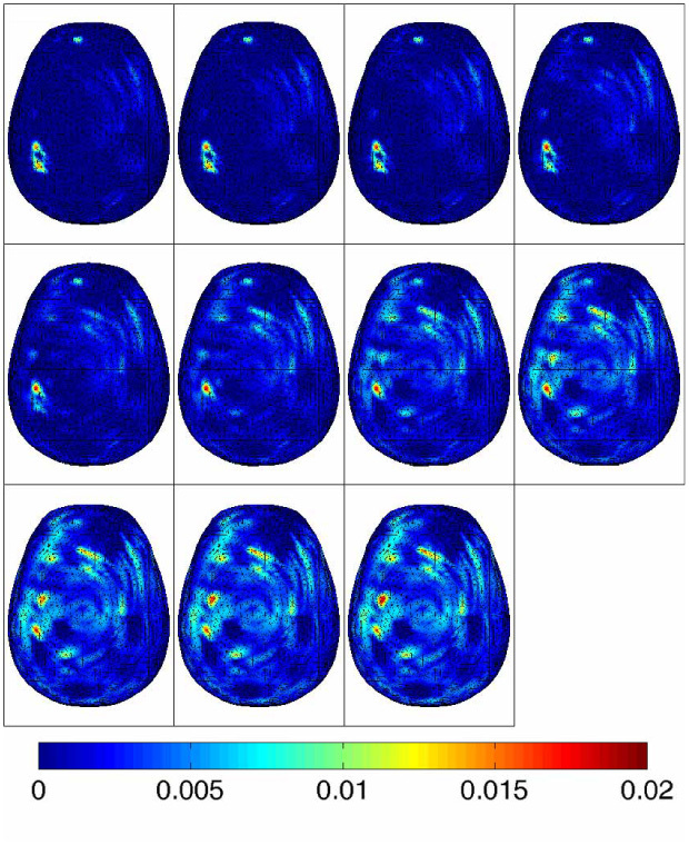
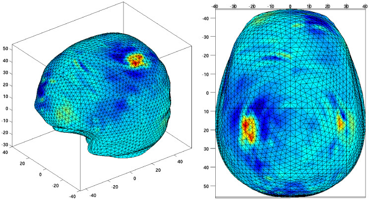
Cortical mapping, also called optical topography is a new medical imaging modality which allows the non-invasive investigation of the outer layers of the cortex. This technique is challenging and the geometry of the subject is very often over-simplified. We aim here to localize activated regions of an anatomically accurate brain. A Boundary Element Method is used for the forward model. The reconstruction of perturbations in the absorption coefficient is demonstrated in a geometrically realistic simulation and in vivo. These results show that diffuse optical imaging of the head can provide reliable activity maps when anatomical data is available.
Keywords: (110.0113) Imaging through turbid media; (110.3200) Inverse scattering; (170.3010) Image reconstruction techniques.
Figures
Similar articles
-
Levels of detail analysis of microwave scattering from human head models for brain stroke detection.PeerJ. 2017 Nov 21;5:e4061. doi: 10.7717/peerj.4061. eCollection 2017. PeerJ. 2017. PMID: 29177115 Free PMC article.
-
Implementation of a phase array diffuse optical tomographic imager.Rev Sci Instrum. 2008 Aug;79(8):084301. doi: 10.1063/1.2963042. Rev Sci Instrum. 2008. PMID: 19044366
-
Combination of boundary element method and finite element method in diffuse optical tomography.IEEE Trans Biomed Eng. 2010 Nov;57(11). doi: 10.1109/TBME.2010.2055868. Epub 2010 Jul 26. IEEE Trans Biomed Eng. 2010. PMID: 20667804
-
Development of volume conductor and source models to localize epileptic foci.J Clin Neurophysiol. 2007 Apr;24(2):101-19. doi: 10.1097/WNP.0b013e318038fb3e. J Clin Neurophysiol. 2007. PMID: 17414966 Review.
-
Review on solving the forward problem in EEG source analysis.J Neuroeng Rehabil. 2007 Nov 30;4:46. doi: 10.1186/1743-0003-4-46. J Neuroeng Rehabil. 2007. PMID: 18053144 Free PMC article. Review.
Cited by
-
High-density diffuse optical tomography for imaging human brain function.Rev Sci Instrum. 2019 May;90(5):051101. doi: 10.1063/1.5086809. Rev Sci Instrum. 2019. PMID: 31153254 Free PMC article.
References
-
- Arridge S. R., “Optical tomography in medical imaging,” Inverse Probl. 15(2), R41–R93 (1999).10.1088/0266-5611/15/2/022 - DOI
LinkOut - more resources
Full Text Sources