Case Reports
. 2011 Jun;50(6):466-77.
doi: 10.1002/gcc.20872. Epub 2011 Mar 15.

Identification of intragenic deletions and duplication in the FLCN gene in Birt-Hogg-Dubé syndrome

Affiliations
Case Reports

Identification of intragenic deletions and duplication in the FLCN gene in Birt-Hogg-Dubé syndrome

Jihane N Benhammou et al. Genes Chromosomes Cancer. 2011 Jun.

Abstract

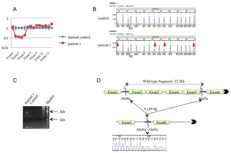
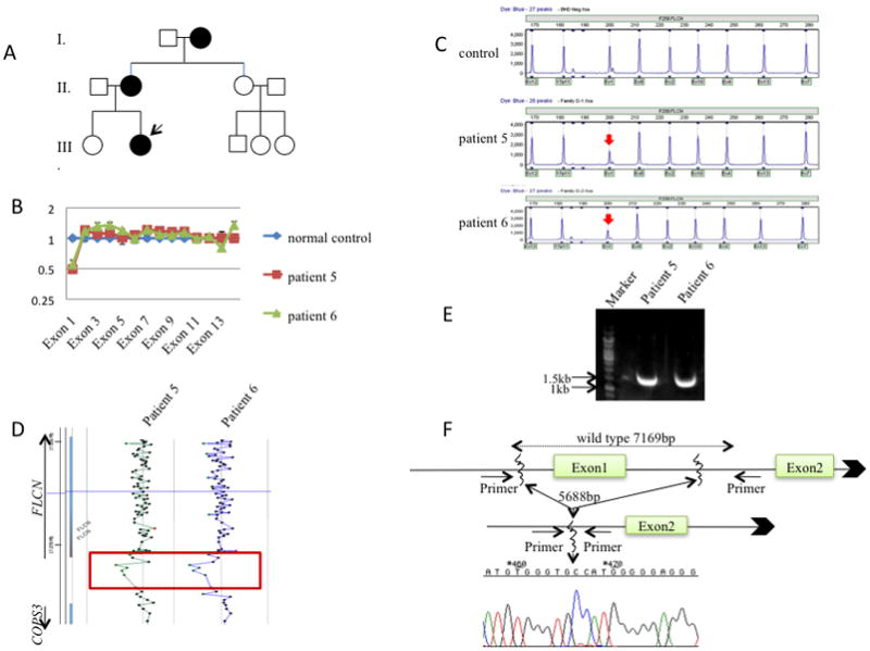
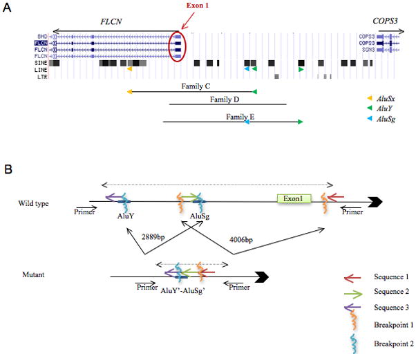
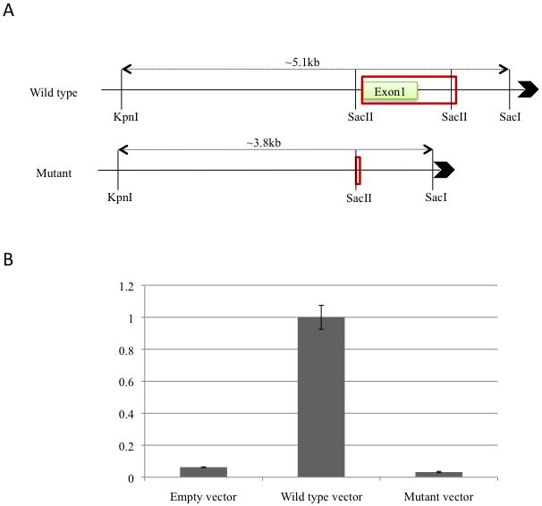
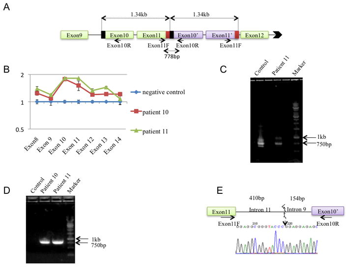
Birt-Hogg-Dubé syndrome (BHDS), caused by germline mutations in the folliculin (FLCN) gene, predisposes individuals to develop fibrofolliculomas, pulmonary cysts, spontaneous pneumothoraces, and kidney cancer. The FLCN mutation detection rate by bidirectional DNA sequencing in the National Cancer Institute BHDS cohort was 88%. To determine if germline FLCN intragenic deletions/duplications were responsible for BHDS in families lacking FLCN sequence alterations, 23 individuals from 15 unrelated families with clinically confirmed BHDS but no sequence variations were analyzed by real-time quantitative PCR (RQ-PCR) using primers for all 14 exons. Multiplex ligation-dependent probe amplification (MLPA) assay and array-based comparative genomic hybridization (aCGH) were utilized to confirm and fine map the rearrangements. Long-range PCR followed by DNA sequencing was used to define the breakpoints. We identified six unique intragenic deletions in nine patients from six different BHDS families including four involving exon 1, one that spanned exons 2-5, and one that encompassed exons 7-14 of FLCN. Four of the six deletion breakpoints were mapped, revealing deletions ranging from 5688 to 9189 bp. In addition, one 1341 bp duplication, which included exons 10 and 11, was identified and mapped. This report confirms that large intragenic FLCN deletions can cause BHDS and documents the first large intragenic FLCN duplication in a BHDS patient. Additionally, we identified a deletion "hot spot" in the 5'-noncoding-exon 1 region that contains the putative FLCN promoter based on a luciferase reporter assay. RQ-PCR, MLPA and aCGH may be used for clinical molecular diagnosis of BHDS in patients who are FLCN mutation-negative by DNA sequencing.


Conflict of interest statement

COMPETING INTERESTS

None.

Figures

Figure 1
Figure 1. Mapping of FLCN exon 2–5 deletion in patient 1 of BHDS Family A.
A. RQ-PCR of genomic DNA from patient 1 demonstrated a heterozygous deletion of exons 2–5. B. MLPA analysis revealed a heterozygous deletion of exons 2–5 of the FLCN gene in patient 1(lower panel) compared with normal control (upper panel). C. LR-PCR of patient 1 DNA generated a smaller mutant fragment of ~3kb not seen in control sample. D. Sequencing of the breakpoint confirmed a deletion of 9189bp. The breakpoint contained sequences from both AluSq and AluSx repeat elements at the 5′- and 3′-boundaries of the deletion.
Figure 2
Figure 2. Mapping of FLCN exon 1 deletions in patients 5 and 6 of BHDS Family D
A. Pedigree of Family D. Arrow indicates proband (patient 6). B. RQ-PCR of DNA from patients 5 (mother) and 6 (proband) demonstrated a heterozygous deletion of exon 1 of the FLCN gene. C. MLPA analysis revealed a heterozygous deletion of exon 1 for patients 5 and 6 (lower panels) compared with normal control (upper panel). D. aCGH results for patients 5 and 6. Red box, deleted region including exon 1. E. LR-PCR of region of interest in DNA from patients 5 and 6 yielded smaller mutant products of ~1.5kb using primers BHDpromoter4 and BHDintron1d. F. Sequencing of mutant PCR products confirmed deletion of 5688bp in both proband and mother, which is not flanked by Alu repeat elements. Large arrowhead indicates 5′-3′ direction.
Figure 3
Figure 3. Genomic location of FLCN exon 1 deletions in BHDS Families C, D and E and map of complex FLCN deletion in BHDS Family E
A. Location of all mapped exon 1 deletions relative to location of known SINE, LINE and LTR repeat elements. The black lines represent the deleted segments in Families C, D and E. All mapped deletions are unique and none involves the adjacent upstream gene, COPS3. Colored triangles, Alu sequences. B. The complex FLCN deletion in Family E most likely resulted from an initial inversion event that involved an exon 1-containing 4006bp sequence flanked by breakpoint 1 boundaries (orange vertical lines), or a 2889bp sequence upstream of exon 1 flanked by breakpoint 2 boundaries containing Alu sequences (blue vertical lines). A subsequent 6645bp deletion event occurred that eliminated exon 1 but retained a 125bp sequence (green arrow) in the reverse orientation. The AluY and AluSg elements that flanked the 2889bp sequence may have mediated one, but not both, of these events. Red arrow, intron 1 sequence; purple arrow, sequence 5′ to deleted sequences. Large arrowhead indicates 5′-3′ direction.
Figure 4
Figure 4. FLCN promoter/exon 1 luciferase reporter assay
A. A wild type FLCN promoter/exon 1 DNA fragment containing a 5149bp region encompassing 4248bp 5′ of exon 1, exon 1 (228bp) and 673bp of intron 1 was inserted into the pGL3 luciferase reporter vector. A mutant FLCN promoter/exon 1 deletion fragment that lacked exon 1, a region commonly deleted in BHDS Families C, D, E, and F, was inserted into the luciferase reporter vector. B. FLCN promoter/exon 1 deletion mutant vector displayed 31 fold less activity than the wild-type FLCN luciferase reporter vector when transfected into HEK293-A cells. Empty vector control is included for comparison. Y-axis, relative activity normalized to wild-type FLCN luciferase reporter activity. Red box, predicted CpG island sequence. Large arrowhead indicates 5′-3′ direction.
Figure 5
Figure 5. Mapping of FLCN exons 10 and 11 duplication identified in BHDS Family G
A. Schematic diagram showing the 1.34kb duplication of exons 10 and 11 of the FLCN gene in tandem with wild-type exons 10 and 11. PCR amplification of patient 11 DNA using exon11QF and exon10QR primers produced a 778bp product that included intron 9 sequence (black box) and intron 11 sequence (red box). B. RQ-PCR of DNA from patients 10 and 11 revealed a heterozygous duplication of exons 10 and 11. C. PCR amplification of FLCN cDNA from patient 11 tumor samples revealed both wild-type and larger mutant products resulting from the duplication event. D. PCR amplification of the exons 11 and 10′ breakpoint in genomic sequence of proband and his affected sister. E. Sequencing of mutant FLCN duplication PCR product defined the breakpoint.

References

    1. Ahvenainen T, Lehtonen HJ, Lehtonen R, Vahteristo P, Aittomaki K, Baynam G, Dommering C, Eng C, Gruber SB, Gronberg H, Harvima R, Herva R, Hietala M, Kujala M, Kaariainen H, Sunde L, Vierimaa O, Pollard PJ, Tomlinson IP, Bjorck E, Aaltonen LA, Launonen V. Mutation screening of fumarate hydratase by multiplex ligation-dependent probe amplification: detection of exonic deletion in a patient with leiomyomatosis and renal cell cancer. Cancer Genet Cytogenet. 2008;183:83–88. - PubMed
    1. Baba M, Hong SB, Sharma N, Warren MB, Nickerson ML, Iwamatsu A, Esposito D, Gillette WK, Hopkins RF, III, Hartley JL, Furihata M, Oishi S, Zhen W, Burke TR, Jr, Linehan WM, Schmidt LS, Zbar B. Folliculin encoded by the BHD gene interacts with a binding protein, FNIP1, and AMPK, and is involved in AMPK and mTOR signaling. Proc Natl Acad Sci U S A. 2006;103:15552–15557. - PMC - PubMed
    1. Birt AR, Hogg GR, Dube WJ. Hereditary multiple fibrofolliculomas with trichodiscomas and acrochordons. Arch Dermatol. 1977;113:1674–1677. - PubMed
    1. Chang YF, Imam JS, Wilkinson MF. The nonsense-mediated decay RNA surveillance pathway. Annu Rev Biochem. 2007;76:51–74. - PubMed
    1. De La Torre C, Ocampo C, Doval IG, Losada A, Cruces MJ. Acrochordons are not a component of the Birt-Hogg-Dube syndrome: does this syndrome exist? Case reports and review of the literature. Am J Dermatopathol. 1999;21:369–374. - PubMed

Publication types