. 2011 Apr;65(4):1125-30.
doi: 10.1002/mrm.22700. Epub 2010 Nov 30.

Phase-sensitive sodium B1 mapping

Affiliations

Phase-sensitive sodium B1 mapping

Steven P Allen et al. Magn Reson Med. 2011 Apr.

Abstract

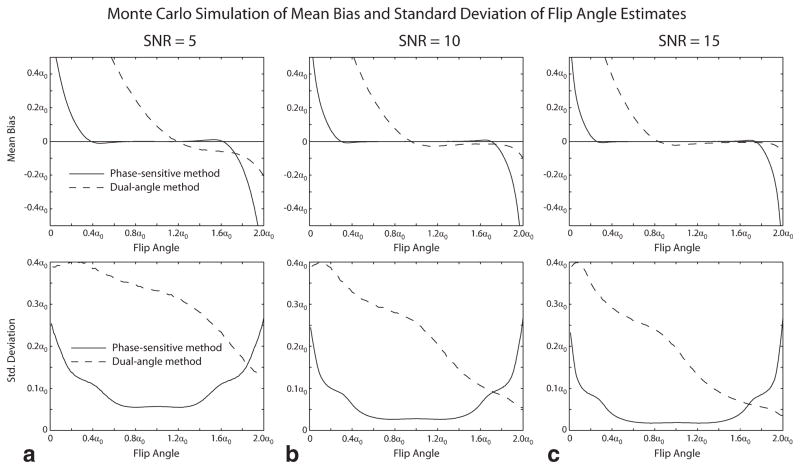
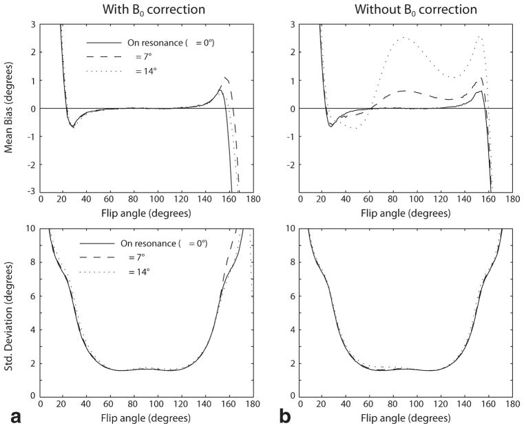
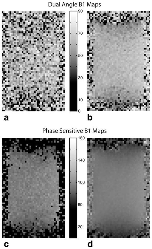
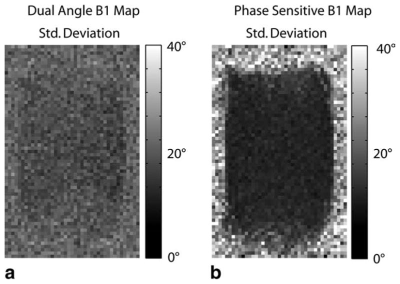
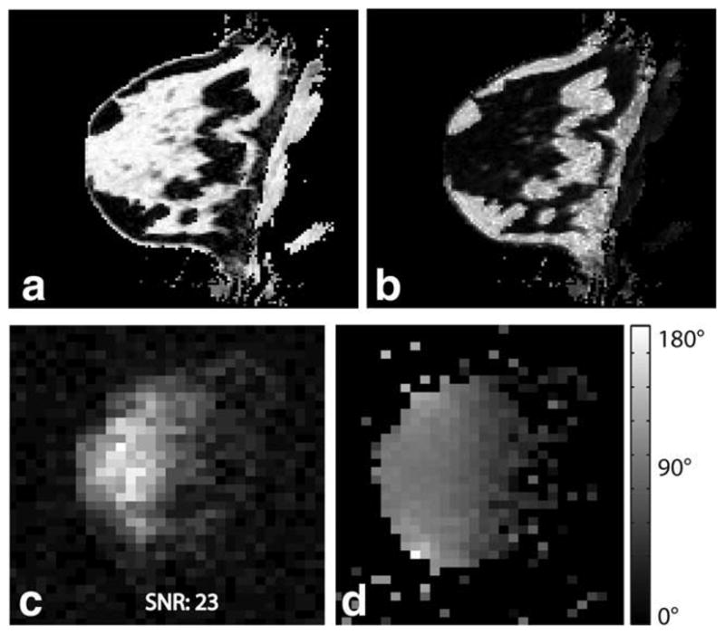
Quantitative sodium MRI requires accurate knowledge of factors affecting the sodium signal. One important determinant of sodium signal level is the transmit B(1) field strength. However, the low signal-to-noise ratio typical of sodium MRI makes accurate B(1) mapping in reasonable scan times challenging. A new phase-sensitive B(1) mapping technique has recently been shown to work better than the widely used dual-angle method in low-signal-to-noise ratio situations and over a broader range of flip angles. In this work, the phase-sensitive B(1) mapping technique is applied to sodium, and its performance compared to the dual-angle method through both simulation and phantom studies. The phase-sensitive method is shown to yield higher quality B(1) maps at low signal-to-noise ratio and greater consistency of measurement than the dual-angle method. An in vivo sodium B(1) map of the human breast is also shown, demonstrating the phase-sensitive method's feasibility for human studies.


Figures

FIG. 1
FIG. 1
Monte Carlo simulation of dual-angle and phase-sensitive B1 mapping methods. The mean bias (upper panels) and standard deviation (lower panels) of the flip angle estimate obtained by each method over a range of actual flip angles are shown for values of system SNR ranging from 5 to 15 (panels ac). The phase-sensitive method is seen to have lower flip angle estimate mean bias and lower standard deviation over most of the measurable range of flip angles.
FIG. 2
FIG. 2
Monte Carlo simulation of effect of off-resonance on the phase-sensitive method. The left panels (a) show the effect of off-resonance on the mean bias and standard deviation of the phase-sensitive flip angle estimate when B0 correction is performed, as described in Ref. . The right panels (b) show the effect of off-resonance if no B0 correction is performed. Off-resonance phase accrual during the RF excitation causes mean bias, but has little effect on the standard deviation of the estimate for the values of off-resonance precession of 7° and 14° shown. Note that these values were informed by the in vivo study shown in Fig. 5, where the maximum off-resonance precession angle across the breast was measured to be about 7°.
FIG. 3
FIG. 3
Dual-angle sodium B1 maps for a single acquisition (a) and 20 averaged acquisitions (b). Phase-sensitive sodium B1 maps for a single acquisition (c) and 20 averaged acquisitions (d). As seen, the phase-sensitive method yields much more robust sodium B1 maps than the dual-angle method. Note that the phase-sensitive maps are centered around a nominal flip angle of 90° vs. 60° for the dual-angle method, reflecting the inherently wider dynamic range of the phase-sensitive method.
FIG. 4
FIG. 4
Standard deviation of the sodium B1 maps for both the dual-angle method (a) and phase-sensitive method (b) as measured across 20 acquisitions. The phase-sensitive B1 mapping method yields a much more consistent measurement in the low-SNR environment typical of sodium imaging.
FIG. 5
FIG. 5
Proton three-point Dixon water (a) and fat (b) image of the breast of a healthy volunteer. The corresponding sodium magnitude image is shown in (c), showing good correlation with breast anatomy (high sodium concentration in fibroglandular tissue and lower concentration in fatty tissue). A 3D sodium B1 map using the phase-sensitive method is shown in (d). The phase-sensitive method yields a high-quality B1 map in vivo despite low sodium image SNR.

References

    1. Borthakur A, Shapiro EM, Beers J, Kudchodkar S, Kneeland JB, Reddy R. Sensitivity of MRI to proteoglycan depletion in cartilage: comparison of sodium and proton MRI. Osteoarthritis Cartilage. 2000;8:288–293. - PubMed
    1. Reddy R, Insko EK, Noyszewski EA, Dandora R, Kneeland JB, Leigh JS. Sodium MRI of human articular cartilage in vivo. Magn Reson Med. 1998;39:697–701. - PubMed
    1. Shapiro EM, Borthakur A, Gougoutas A, Reddy R. 23Na MRI accurately measures fixed charge density in articular cartilage. Magn Reson Med. 2002;47:284–291. - PMC - PubMed
    1. Wheaton AJ, Borthakur A, Shapiro EM, Regatte RR, Akella SV, Kneeland JB, Reddy R. Proteoglycan loss in human knee cartilage: quantitation with sodium MR imaging—feasibility study. Radiology. 2004;231:900–905. - PubMed
    1. Ouwerkerk R, Bleich KB, Gillen JS, Pomper MG, Bottomley PA. Tissue sodium concentration in human brain tumors as measured with 23Na MR imaging. Radiology. 2003;227:529–537. - PubMed

Publication types