

Nanoporous elements in microfluidics for multiscale manipulation of bioparticles
- PMID: 21413145
- PMCID: PMC3141316
- DOI: 10.1002/smll.201002076
Nanoporous elements in microfluidics for multiscale manipulation of bioparticles
Abstract
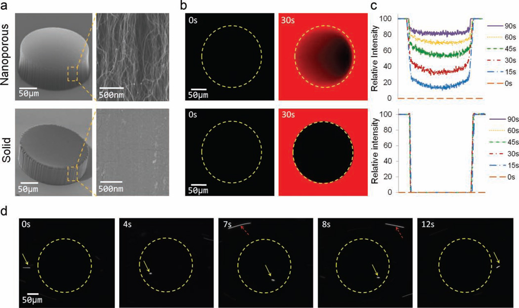
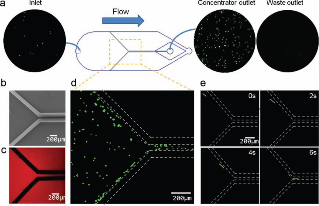
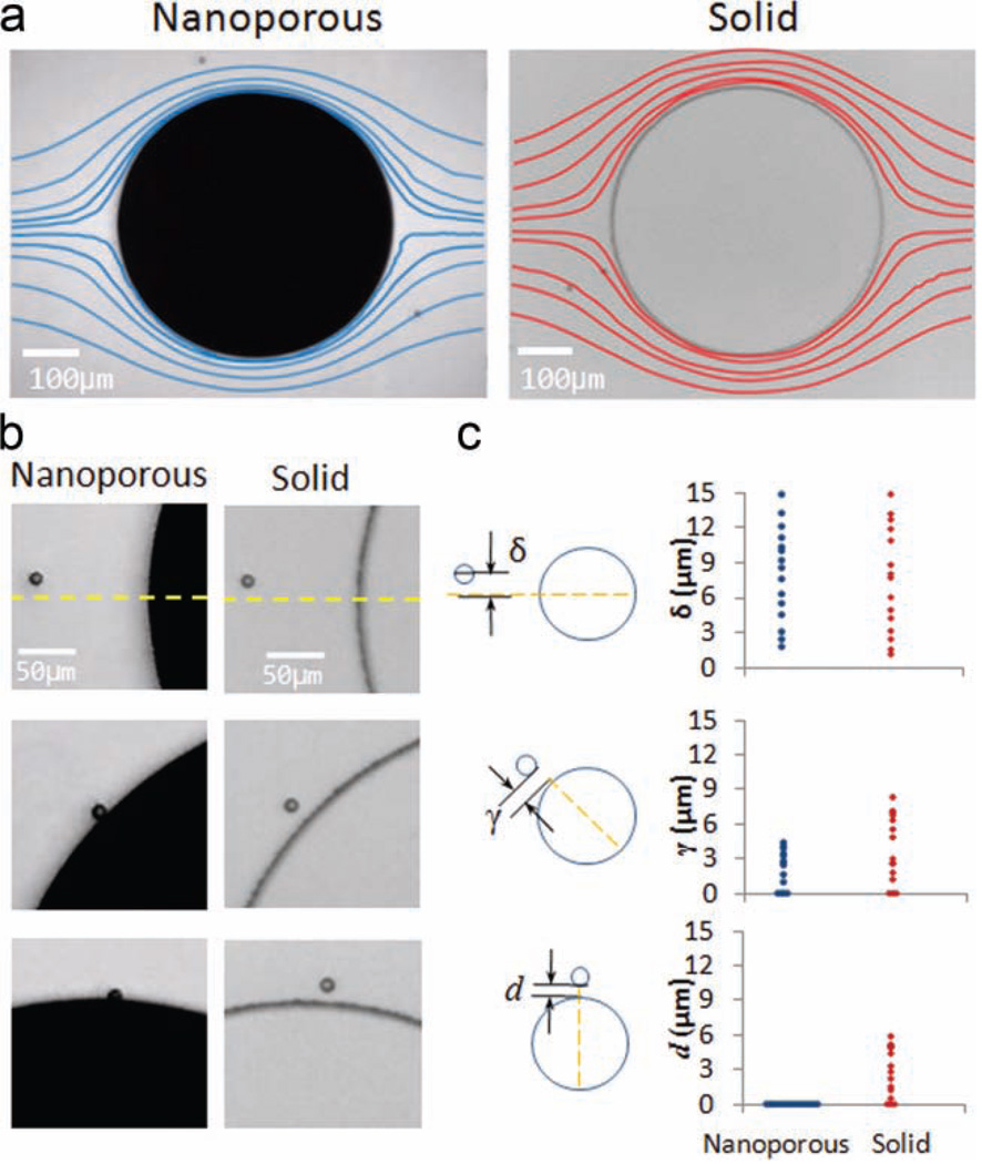
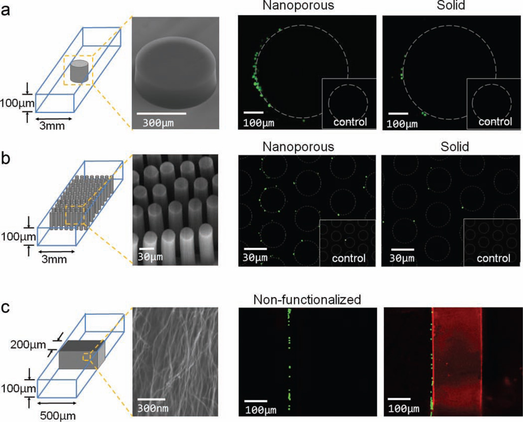
Solid materials, such as silicon, glass, and polymers, dominate as structural elements in microsystems including microfluidics. Porous elements have been limited to membranes sandwiched between microchannel layers or polymer monoliths. This paper reports the use of micropatterned carbon-nanotube forests confined inside microfluidic channels for mechanically and/or chemically capturing particles ranging over three orders of magnitude in size. Nanoparticles below the internanotube spacing (80 nm) of the forest can penetrate inside the forest and interact with the large surface area created by individual nanotubes. For larger particles (>80 nm), the ultrahigh porosity of the nanotube elements reduces the fluid boundary layer and enhances particle-structure interactions on the outer surface of the patterned nanoporous elements. Specific biomolecular recognition is demonstrated using cells (≈10 μm), bacteria (≈1 μm), and viral-sized particles (≈40 nm) using both effects. This technology can provide unprecedented control of bioseparation processes to access bioparticles of interest, opening new pathways for both research and point-of-care diagnostics.
Copyright © 2011 WILEY-VCH Verlag GmbH & Co. KGaA, Weinheim.
Figures
Similar articles
-
Monolithic integration of well-ordered nanoporous structures in the microfluidic channels for bioseparation.J Chromatogr A. 2007 Aug 31;1162(2):175-9. doi: 10.1016/j.chroma.2007.06.037. Epub 2007 Jun 27. J Chromatogr A. 2007. PMID: 17628581
-
Digital microfluidics and delivery of molecular payloads with magnetic porous silicon chaperones.Dalton Trans. 2008 Feb 14;(6):721-30. doi: 10.1039/b714594b. Epub 2007 Nov 16. Dalton Trans. 2008. PMID: 18239825
-
Microfluidics for pharmaceutical nanoparticle fabrication: The truth and the myth.Int J Pharm. 2020 Jun 30;584:119408. doi: 10.1016/j.ijpharm.2020.119408. Epub 2020 May 12. Int J Pharm. 2020. PMID: 32407942 Review.
-
Formation of self-organized nanoporous anodic oxide from metallic gallium.Langmuir. 2012 Sep 25;28(38):13705-11. doi: 10.1021/la302672a. Epub 2012 Sep 10. Langmuir. 2012. PMID: 22934571
-
Methods of Generating Dielectrophoretic Force for Microfluidic Manipulation of Bioparticles.ACS Biomater Sci Eng. 2021 Jun 14;7(6):2043-2063. doi: 10.1021/acsbiomaterials.1c00083. Epub 2021 Apr 19. ACS Biomater Sci Eng. 2021. PMID: 33871975 Free PMC article. Review.
Cited by
-
Enhanced Isolation and Release of Circulating Tumor Cells Using Nanoparticle Binding and Ligand Exchange in a Microfluidic Chip.J Am Chem Soc. 2017 Feb 22;139(7):2741-2749. doi: 10.1021/jacs.6b12236. Epub 2017 Feb 9. J Am Chem Soc. 2017. PMID: 28133963 Free PMC article.
-
A carbon nanotube integrated microfluidic device for blood plasma extraction.Sci Rep. 2018 Sep 11;8(1):13623. doi: 10.1038/s41598-018-31810-x. Sci Rep. 2018. PMID: 30206295 Free PMC article.
-
Nanotechnology for enrichment and detection of circulating tumor cells.Nanomedicine (Lond). 2015 Jul;10(12):1973-90. doi: 10.2217/nnm.15.32. Nanomedicine (Lond). 2015. PMID: 26139129 Free PMC article. Review.
-
Biporous silica nanostructure-induced nanovortex in microfluidics for nucleic acid enrichment, isolation, and PCR-free detection.Nat Commun. 2024 Feb 14;15(1):1366. doi: 10.1038/s41467-024-45467-w. Nat Commun. 2024. PMID: 38355558 Free PMC article.
-
Size-based hydrodynamic rare tumor cell separation in curved microfluidic channels.Biomicrofluidics. 2013 Jan 7;7(1):11802. doi: 10.1063/1.4774311. eCollection 2013. Biomicrofluidics. 2013. PMID: 24396523 Free PMC article.
References
Publication types
MeSH terms
Grants and funding
LinkOut - more resources
Full Text Sources
Other Literature Sources
