

doi: 10.1039/c005374k.
Multiphoton microscopy of transdermal quantum dot delivery using two photon polymerization-fabricated polymer microneedles
Affiliations
- PMID: 21413181
- PMCID: PMC3060378
- DOI: 10.1039/c005374k
Item in Clipboard
Multiphoton microscopy of transdermal quantum dot delivery using two photon polymerization-fabricated polymer microneedles
Faraday Discuss.
2011.
Abstract
Due to their ability to serve as fluorophores and drug delivery vehicles, quantum dots are a powerful tool for theranostics-based clinical applications. In this study, microneedle devices for transdermal drug delivery were fabricated by means of two-photon polymerization of an acrylate-based polymer. We examined proliferation of cells on this polymer using neonatal human epidermal keratinocytes and human dermal fibroblasts. The microneedle device was used to inject quantum dots into porcine skin; imaging of the quantum dots was performed using multiphoton microscopy.
Figures

a) Schematic of the two photon polymerization system. Femtosecond laser pulses pass through a beam expander and a filter wheel before being focused on the objective. Scanning mirrors control the location of the laser spot in the focal plane. Polymerization of the resin occurs at the focal point. A charge-coupled device camera behind a dichroic mirror is used for visualizing the two photon polymerization process. b) Schematic of multiphoton microscopy system. Femtosecond laser pulses pass through the objective before being focused on the specimen, where excitation occurs at the focal point. Scanning mirrors control the location of the laser spot in the focal plane. Dichroic mirrors are used to separate excitation and emission light and to separate emitted light into two channels.

a) Input computer-aided design drawing of hollow microneedles processed via two photon polymerization. b) Diagram of hollow microneedle device fabrication by means of two photon polymerization.

Fourier transform infrared spectrum of e-shell 300 acrylate-based polymer.

MTT viability of neonatal human epidermal keratinocytes and human dermal fibroblasts on e-shell 300 acrylate-based polymer, polystyrene well plate, and glass is shown. Error bars indicate standard error of the mean.

Scanning electron microscopy image obtained at 45° tilt of solid e-shell 300 microneedles, which were created by means of two photon polymerization. Mark speeds (from front to back) of 250, 50, 100, 150, and 200 are shown.
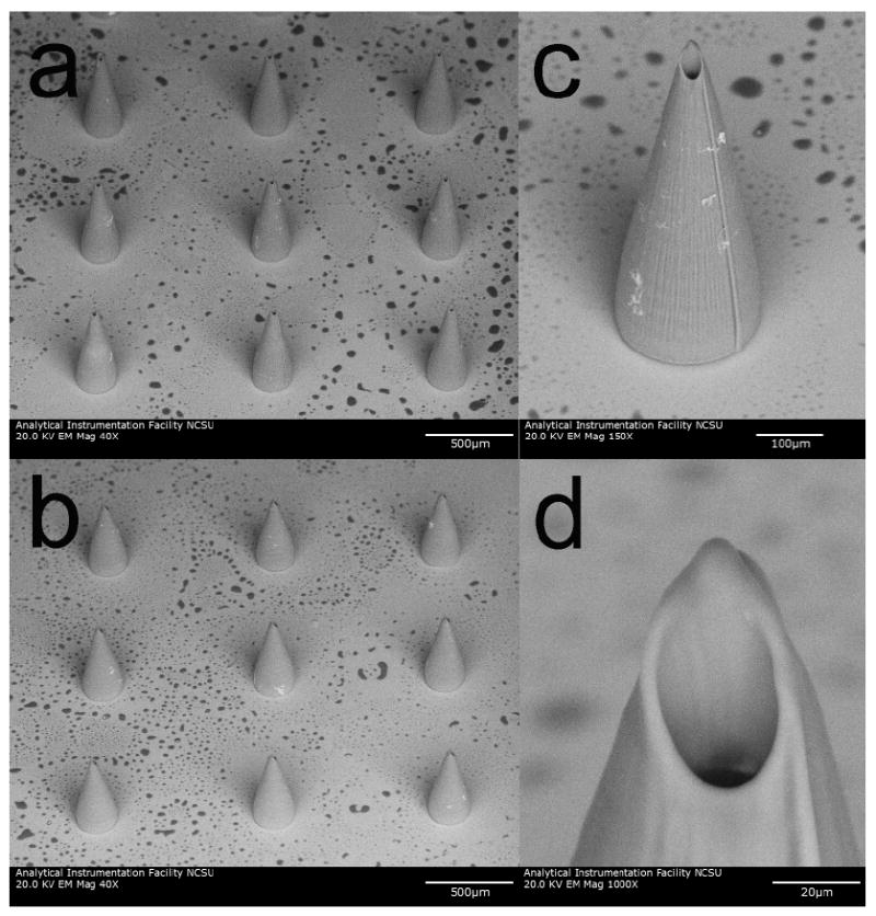
Scanning electron microscopy images obtained at 45° tilt of e-shell 300 hollow microneedles on glass substrates, which were produced using two photon polymerization. a) Image of 614 +/- 12 μm long microneedle array. b) Image of 710 +/- 10 μm long microneedle array. c) Image of 710+/- 10 μm long individual microneedle. d) Image of 710+/- 10 μm long individual microneedle. The base diameter of these microneedles is 226 +/- 5 μm. Dimensions are shown as average +/- standard deviation.

Scanning electron microscopy images of e-shell 300 hollow microneedle array, which was produced using two photon polymerization. a) Image of microneedle array obtained at 45° tilt. b) Image of individual microneedle obtained at 45° tilt. c) Image of individual microneedle obtained at 0° tilt. The length and base diameters of these microneedles are 508 +/- 33 μm and 212 +/- 3 μm, respectively. Dimensions are shown as average +/- standard deviation.
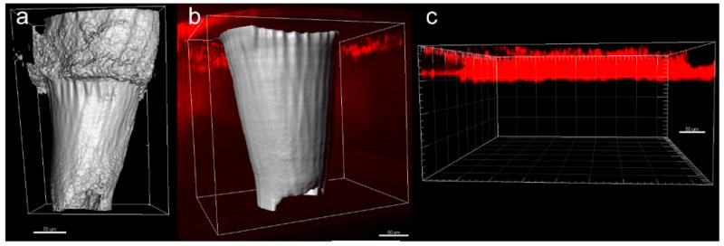
Multiphoton microscopy images of quantum dot injection via two photon polymerization-fabricated e-shell 300 microneedle array as well as via topical application. The microneedles are presented as surface renderings (in gray) and the quantum dots are presented as maximum projections (in red). a) Microneedle in porcine skin prior to quantum dot injection. b) Microneedle in porcine skin after quantum dot injection. A broad distribution of the quantum dots in the deep epidermis and dermis was observed. c) Quantum dots topically applied to porcine skin. The topically applied quantum dots exhibited poor penetration and remained in the topmost 50 μm region of the epidermis.
Similar articles
-
Fabrication of microneedles using two photon polymerization for transdermal delivery of nanomaterials.J Nanosci Nanotechnol. 2010 Oct;10(10):6305-12. doi: 10.1166/jnn.2010.2636. J Nanosci Nanotechnol. 2010. PMID: 21137723
-
Tapered conical polymer microneedles fabricated using an integrated lens technique for transdermal drug delivery.IEEE Trans Biomed Eng. 2007 May;54(5):903-13. doi: 10.1109/TBME.2006.889173. IEEE Trans Biomed Eng. 2007. PMID: 17518288
-
Ex vivo evaluation of a microneedle array device for transdermal application.Int J Pharm. 2015 Dec 30;496(2):351-9. doi: 10.1016/j.ijpharm.2015.09.070. Epub 2015 Oct 21. Int J Pharm. 2015. PMID: 26453791
-
A key role by polymers in microneedle technology: a new era.Drug Dev Ind Pharm. 2021 Nov;47(11):1713-1732. doi: 10.1080/03639045.2022.2058531. Epub 2022 Apr 6. Drug Dev Ind Pharm. 2021. PMID: 35332822 Review.
-
Current trends in polymer microneedle for transdermal drug delivery.Int J Pharm. 2020 Sep 25;587:119673. doi: 10.1016/j.ijpharm.2020.119673. Epub 2020 Jul 30. Int J Pharm. 2020. PMID: 32739388 Free PMC article. Review.
Cited by
-
Progress in Microneedle-Mediated Protein Delivery.J Clin Med. 2020 Feb 17;9(2):542. doi: 10.3390/jcm9020542. J Clin Med. 2020. PMID: 32079212 Free PMC article. Review.
-
Two-Photon Polymerisation 3D Printing of Microneedle Array Templates with Versatile Designs: Application in the Development of Polymeric Drug Delivery Systems.Pharm Res. 2020 Aug 27;37(9):174. doi: 10.1007/s11095-020-02887-9. Pharm Res. 2020. PMID: 32856172 Free PMC article.
-
Digital Manufacturing for Microfluidics.Annu Rev Biomed Eng. 2019 Jun 4;21:325-364. doi: 10.1146/annurev-bioeng-092618-020341. Annu Rev Biomed Eng. 2019. PMID: 31167099 Free PMC article. Review.
-
Revolutionizing Therapeutic Delivery with Microneedle Technology for Tumor Treatment.Pharmaceutics. 2022 Dec 21;15(1):14. doi: 10.3390/pharmaceutics15010014. Pharmaceutics. 2022. PMID: 36678643 Free PMC article. Review.
-
The Effects of Geometry on Skin Penetration and Failure of Polymer Microneedles.J Adhes Sci Technol. 2013 Feb 1;27(3):227-243. doi: 10.1080/01694243.2012.705101. Epub 2012 Aug 6. J Adhes Sci Technol. 2013. PMID: 23543070 Free PMC article.
References
-
- Warner S. Scientist. 2004;18:38–39.
MeSH terms
Substances
Grants and funding
LinkOut - more resources
Full Text Sources
Other Literature Sources