Review
. 2011 May 20;585(10):1412-26.
doi: 10.1016/j.febslet.2011.03.021. Epub 2011 Mar 21.

Circadian disruption and SCN control of energy metabolism

Affiliations
Review

Circadian disruption and SCN control of energy metabolism

Andries Kalsbeek et al. FEBS Lett. .

Abstract

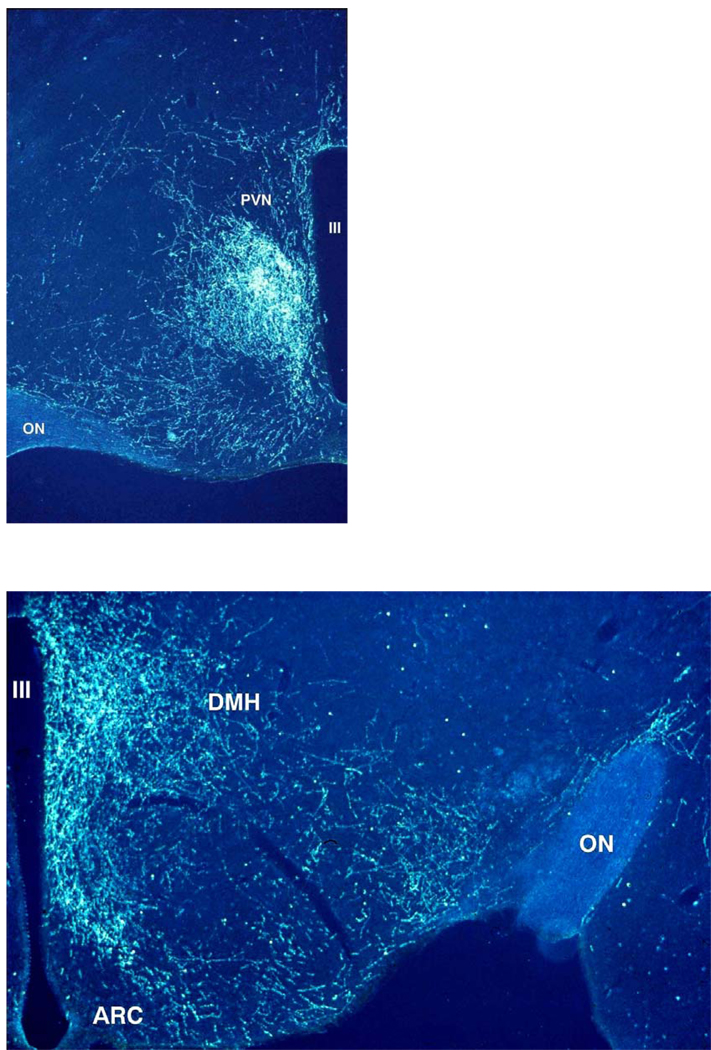
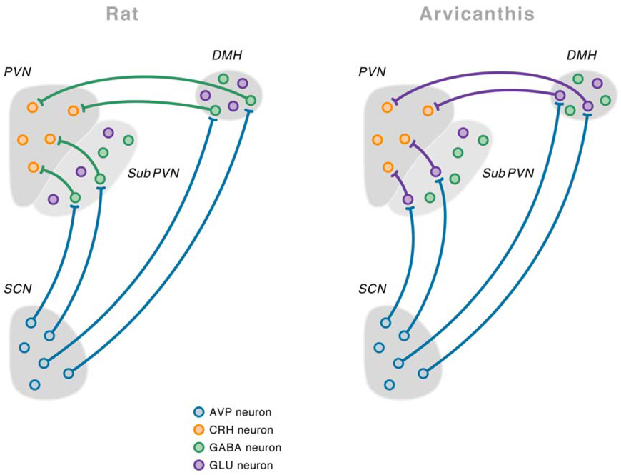
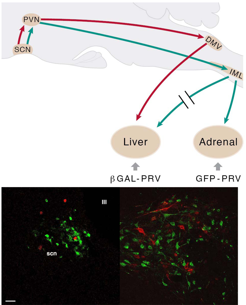
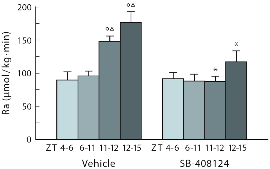
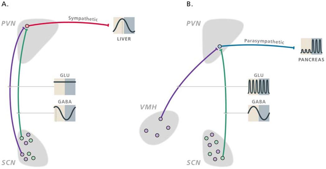
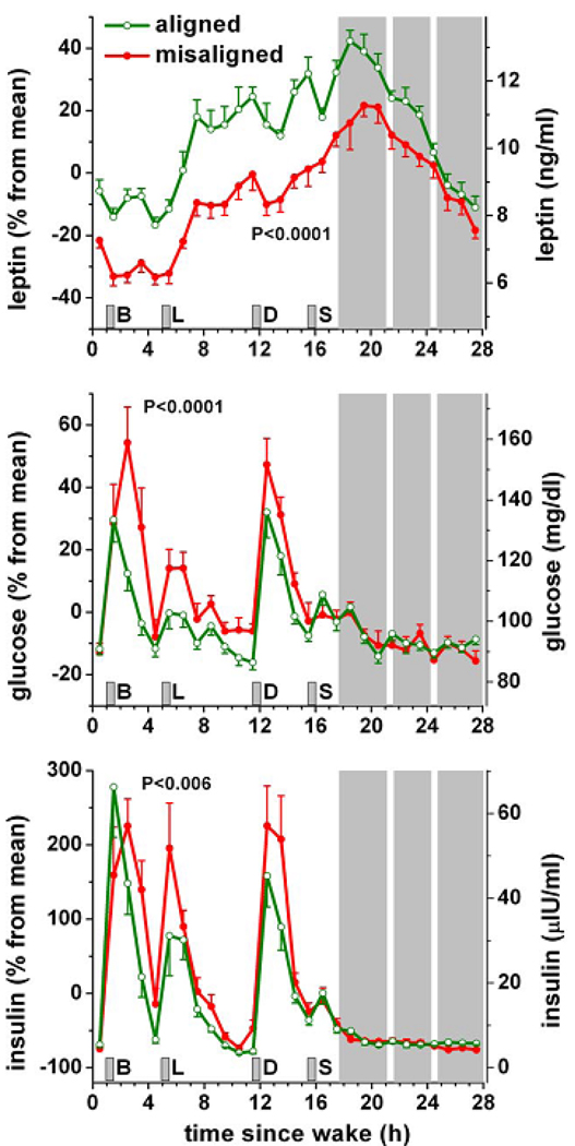
In this review we first present the anatomical pathways used by the suprachiasmatic nuclei to enforce its rhythmicity onto the body, especially its energy homeostatic system. The experimental data show that by activating the orexin system at the start of the active phase, the biological clock not only ensures that we wake up on time, but also that our glucose metabolism and cardiovascular system are prepared for increased activity. The drawback of such a highly integrated system, however, becomes visible when our daily lives are not fully synchronized with the environment. Thus, in addition to increased physical activity and decreased intake of high-energy food, also a well-lighted and fully resonating biological clock may help to withstand the increasing "diabetogenic" pressure of today's 24/7 society.


Figures

Fig.1
Fig.1. Darkfield illustration of SCN projections into the hypothalamic paraventricular (PVN), dorsomedial (DMH) and ventromedial (VMH) hypothalamus
After the iontophoretic injection of Phaseolus vulgaris leucoagglutinin (PHA-L) into the SCN a large concentration of labelled fibers can be observed in the area just ventral to the PVN (upper panel), also known as the sub-paraventricular zone or the subPVN. Less dense but also clearly innervated in the upper panel are the periventricular part and the dorsomedial part of the PVN, but note the almost complete lack of labelled fibers in the parvicellular and magnocellular parts of the PVN. Caudal to the PVN (lower panel) a dense terminal field of SCN fibers can be observed in the DMH. In the lower panel also a clear concentration of PHA-L labelled fibers is present in the area between the arcuate nucleus (ARC) and the VMH, as well as the area lateral to the VMH. The third ventricle (III) is on the right side (upper panel) or left side (lower panel).
Fig.2
Fig.2. Mean firing frequency of vasopressin-positive, vasopressin-negative, and non-characterized neurons analyzed in the in vitro dorsal SCN of the rat
The firing frequency was determined in hypothalamic slices at either CT5–8 or CT14–17. All groups showed a significant effect of diurnal timing. However, the difference was only two-fold in the vasopressin-negative neurons, whereas it amounted to five- or six-fold in the vasopressin-positive neurons. Especially the nocturnal firing frequency rate of vasopressin-positive neurons was remarkably lower as compared to the other groups. With permission from (Buijs et al., 2006). ***, p<0.001; *, # p<0.05
Fig.3
Fig.3. Detailed anatomical scheme of demonstrated and putative* connections of the suprachiasmatic nucleus (SCN) in the rat and Arvicanthis ansorgei brain to explain the opposite effects of vasopressin on the HPA axis in these two species
Vasopressin is released during the light period, both in the nocturnal rat and the diurnal Arvicanthis ansorgei. In rats vasopressin release during the light period will inhibit the corticotropin-releasing hormone (CRH)-containing neurons in the paraventricular nucleus of the hypothalamus (PVN) by contacting γ-aminobutyric acid (GABA)ergic interneurons in the subPVN and dorsomedial nucleus of the hypothalamus (DMH). On the other hand, in the Arvicanthis ansorgei, vasopressin release during the light period will stimulate CRH-containing neurons because it acts on the glutamatergic, instead of GABAergic, interneurons in the subPVN and DMH. *Only the projections from the subPVN and DMH to the PVN in the Arvicanthis ansorgei have not been formerly confirmed by tracing experiments. With permission from (Kalsbeek et al., 2008).
Fig.4
Fig.4. Spatial and temporal distribution of Per1 gene expression in the SCN, throughout the L/D-cycle
Per1 gene expression is shown for ad libitum fed animals (AL), for ad libitum fed animals that had been phase-shifted (8-hours) 5 days earlier (ALs), and for animals on a restricted-feeding protocol that had been phase-shifted 5 days earlier (RFs). Per1 gene expression was revealed by the non-radioactive immuno-in situ hybridization technique (with the help of Dr. Hugues Dardente, in the laboratory of Dr. Paul Pévet). Gray and black bars on the left side of the figures indicate light and dark portions of the L/D-cycle. Animals on restricted feeding (RF) had daily access to food from ZT4–6. The 8-h phase-advance of the L/D-cycle was realized by shutting down the light at ZT4. The RF animals had been on the RF protocol for 4 weeks before the phase-shift. From the first day of the shift, the animals were provided with food ad libitum. Starting at ZT10 on day 5 after the shift (D5) and ZT2 for the non-shifted groups, the animals of the 4 groups were maintained in constant darkness under red-dim-light until the end of the experiment. The animals of the AL, ALs, and RFs groups were perfused every 4-hours at six different time points starting at CTs14 on D5 (CTs14 indicating CT14 after the shift and corresponding to ZT6 in non-shifted time). For further details see the thesis of Stephanie Perreau-Lenz (2004).
Fig.5
Fig.5. The suprachiasmatic nucleus (SCN) balances sympathetic and parasympathetic output to peripheral organs through separate pre-autonomic neurons
In the upper panel the experimental setup used to examine the possible separation of sympathetic and parasympathetic pre-autonomic neurons in the hypothalamus is indicated in a schematic drawing of a sagittal section of the rat brain. B-galactoside PRV (βGAL-PRV) was injected into the sympathetic denervated liver, forcing the virus to infect the brain via the vagus nerve (red lines); simultaneously the pre-sympathetic neurons were labelled by an injection of green fluorescent protein (GFP)-PRV into the adrenal (green lines). After the labelling of the first-order neurons in the brainstem and spinal cord, this approach resulted in separate pre-parasympathetic and sympathetic neurons in the PVN (second order), followed by a similar separation of the third-order neurons in the SCN. In the lower panels transverse sections of the hypothalamus in the region of the PVN (left) and SCN (right) show a perfect separation of pre-parasympathetic βGAL-PRV (red) and pre-sympathetic GFP-PRV (green) labelled neurons, as there are no yellow (i.e., double-labelled) neurons. Scale bar = 25 µm. With permission adapted from (Buijs et al., 2003).
Fig.6
Fig.6. Orexin release is necessary for the endogenous rise in glucose appearance at dusk in nocturnal animals
The endogenous rise in glucose appearance (Ra) during ICV vehicle administration is shown in the left panel. ICV administration of the orexin antagonist, during the latter part of the light period, prevents the endogenous rise of Ra before the onset of darkness (ZT11–ZT12), and during the onset of the dark period (ZT12–ZT15) (right panel). The orexin antagonist SB-408124 was administered ICV from ZT4 – ZT12. IV administration of the orexin antagonist had no effect on glucose appearance. Food intake was not changed by ICV orexin. O and Δ, P<0.05 compared with ZT4–6 and ZT6–11, respectively. *, P<0.05 compared with comparable time points in the vehicle group.
Fig.7
Fig.7. Schematic presentation of the daily activity pattern of hypothalamic populations of GABAergic and glutamatergic neurons implicated in the autonomic control of the daily rhythms in hepatic glucose production (A) and feeding-induced insulin release (B)
The sympathetic and parasympathetic pre-autonomic neurons are inhibited by a rhythmic GABAergic input (green dots and lines) from the SCN that is mainly active during the light period. Sympathetic pre-autonomic neurons are stimulated by glutamatergic inputs (purple dots and lines) from the SCN (A), whereas parasympathetic pre-autonomic neurons are stimulated by glutamatergic inputs from the VMH (B). The glutamatergic stimulation only translates in activity of the pre-autonomic neurons when the inhibitory input from the SCN is absent. The control of the sympathetic pre-autonomic neurons that are involved in the control of the daily melatonin rhythm looks very much similar to that of hepatic glucose production, the only difference being a slight phase delay of the activity of GABAergic SCN neurons that innervate the “pineal-dedicated” pre-autonomic neurons. With permission from (Kalsbeek et al., 2008).
Fig.8
Fig.8. Consequences of circadian misalignment on metabolic and endocrine function
Data from 10 healthy volunteers are plotted according to time-since-wake, during normal circadian alignment (open green symbols; scheduled awakening at habitual wake time) and during circadian misalignment (filled red symbols; scheduled awakening 12 h out of phase from habitual wake time). P-values, statistical significance for effect of misalignment; gray area, scheduled sleep episode; short vertical gray bars, meal times; B, breakfast; L, lunch; D, dinner; S, snack. Reproduced with permission from Scheer et al. (2009).

References

    1. Moore RM. Entrainment pathways and the functional organization of the circadian system. Prog. Brain Res. 1996;111:103–119. - PubMed
    1. Challet E, Pévet P. Interactions between photic and nonphotic stimuli to synchronize the master circadian clock in mammals. Front. Biosci. 2003;8:S246–S257. - PubMed
    1. Takahashi JS, Hong HK, Ko CH, McDearmon EL. The genetics of mammalian circadian order and disorder: implications for physiology and disease. Nat. Rev. Genet. 2008;9:764–775. - PMC - PubMed
    1. Jin XW, Shearman LP, Weaver DR, Zylka MJ, De Vries GJ, Reppert SM. A molecular mechanism regulating rhythmic output from the suprachiasmatic circadian clock. Cell. 1999;96:57–68. - PubMed
    1. Hahm SH, Eiden LE. Five discrete cis-active domains direct cell type-specific transcription of the vasoactive intestinal peptide (VIP) gene. J. Biol. Chem. 1998;273:17086–17094. - PubMed

Publication types

MeSH terms