Chronic assessment of diaphragm muscle EMG activity across motor behaviors
- PMID: 21414423
- PMCID: PMC3103648
- DOI: 10.1016/j.resp.2011.03.011
Chronic assessment of diaphragm muscle EMG activity across motor behaviors
Abstract
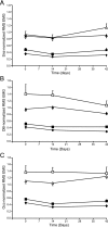
The diaphragm muscle is the main inspiratory muscle in mammals. Quantitative analyses documenting the reliability of chronic diaphragm EMG recordings are lacking. Assessment of ventilatory and non-ventilatory motor behaviors may facilitate evaluating diaphragm EMG activity over time. We hypothesized that normalization of diaphragm EMG amplitude across behaviors provides stable and reliable parameters for longitudinal assessments of diaphragm activity. We found that diaphragm EMG activity shows substantial intra-animal variability over 6 weeks, with coefficient of variation (CV) for different behaviors ∼ 29-42%. Normalization of diaphragm EMG activity to near maximal behaviors (e.g., deep breathing) reduced intra-animal variability over time (CV ∼ 22-29%). Plethysmographic measurements of eupneic ventilation were also stable over 6 weeks (CV ∼ 13% for minute ventilation). Thus, stable and reliable measurements of diaphragm EMG activity can be obtained longitudinally using chronically implanted electrodes by examining multiple motor behaviors. By quantitatively determining the reliability of longitudinal diaphragm EMG analyses, we provide an important tool for evaluating the progression of diseases or injuries that impair ventilation.
Copyright © 2011 Elsevier B.V. All rights reserved.
Figures


Similar articles
-
Diaphragm muscle activity across respiratory motor behaviors in awake and lightly anesthetized rats.J Appl Physiol (1985). 2018 Apr 1;124(4):915-922. doi: 10.1152/japplphysiol.01004.2017. Epub 2018 Jan 4. J Appl Physiol (1985). 2018. PMID: 29357493 Free PMC article.
-
Non-stationarity and power spectral shifts in EMG activity reflect motor unit recruitment in rat diaphragm muscle.Respir Physiol Neurobiol. 2013 Jan 15;185(2):400-9. doi: 10.1016/j.resp.2012.08.020. Epub 2012 Sep 7. Respir Physiol Neurobiol. 2013. PMID: 22986086 Free PMC article.
-
Diaphragm motor unit recruitment in rats.Respir Physiol Neurobiol. 2010 Aug 31;173(1):101-6. doi: 10.1016/j.resp.2010.07.001. Epub 2010 Jul 8. Respir Physiol Neurobiol. 2010. PMID: 20620243 Free PMC article.
-
Phrenic motor unit recruitment during ventilatory and non-ventilatory behaviors.Respir Physiol Neurobiol. 2011 Oct 15;179(1):57-63. doi: 10.1016/j.resp.2011.06.028. Epub 2011 Jul 6. Respir Physiol Neurobiol. 2011. PMID: 21763470 Free PMC article. Review.
-
Diaphragm electromyography using an oesophageal catheter: current concepts.Clin Sci (Lond). 2008 Oct;115(8):233-44. doi: 10.1042/CS20070348. Clin Sci (Lond). 2008. PMID: 18782085 Review.
Cited by
-
Localized delivery of brain-derived neurotrophic factor-expressing mesenchymal stem cells enhances functional recovery following cervical spinal cord injury.J Neurotrauma. 2015 Feb 1;32(3):185-93. doi: 10.1089/neu.2014.3464. Epub 2014 Dec 10. J Neurotrauma. 2015. PMID: 25093762 Free PMC article.
-
V2a neurons restore diaphragm function in mice following spinal cord injury.Proc Natl Acad Sci U S A. 2024 Mar 12;121(11):e2313594121. doi: 10.1073/pnas.2313594121. Epub 2024 Mar 5. Proc Natl Acad Sci U S A. 2024. PMID: 38442182 Free PMC article.
-
Functional recovery after cervical spinal cord injury: Role of neurotrophin and glutamatergic signaling in phrenic motoneurons.Respir Physiol Neurobiol. 2016 Jun;226:128-36. doi: 10.1016/j.resp.2015.10.009. Epub 2015 Oct 23. Respir Physiol Neurobiol. 2016. PMID: 26506253 Free PMC article. Review.
-
Plasticity Induced Recovery of Breathing Occurs at Chronic Stages after Cervical Contusion.J Neurotrauma. 2019 Jun 15;36(12):1985-1999. doi: 10.1089/neu.2018.6186. Epub 2019 Feb 19. J Neurotrauma. 2019. PMID: 30565484 Free PMC article.
-
Neuromotor control of spontaneous quiet breathing in awake rats evaluated by assessments of diaphragm EMG stationarity.J Neurophysiol. 2023 Nov 1;130(5):1344-1357. doi: 10.1152/jn.00267.2023. Epub 2023 Oct 25. J Neurophysiol. 2023. PMID: 37877195 Free PMC article.
References
-
- Alford EK, Roy RR, Hodgson JA, Edgerton VR. Electromyography of rat soleus, medial gastrocnemius, and tibialis anterior during hind limb suspension. Exp. Neurol. 1987;96:635–649. - PubMed
-
- Bell HJ, Haouzi P. The hypoxia-induced facilitation of augmented breaths is suppressed by the common effect of carbonic anhydrase inhibition. Respir. Physiol. Neurobiol. 2010;171:201–211. - PubMed
-
- Blewett C, Elder GC. Quantitative EMG analysis in soleus and plantaris during hindlimb suspension and recovery. J. Appl. Physiol. 1993;74:2057–2066. - PubMed
-
- Bolcskei K, Tekus V, Dezsi L, Szolcsanyi J, Petho G. Antinociceptive desensitizing actions of TRPV1 receptor agonists capsaicin, resiniferatoxin and N-oleoyldopamine as measured by determination of the noxious heat and cold thresholds in the rat. Eur. J. Pain. 2010;14:480–486. - PubMed
-
- Chang FC, Harper RM. A procedure for chronic recording of diaphragmatic electromyographic activity. Brain Res. Bull. 1989;22:561–563. - PubMed
Publication types
MeSH terms
Grants and funding
LinkOut - more resources
Full Text Sources

