. 2011 Feb 23;31(8):2895-905.
doi: 10.1523/JNEUROSCI.5338-10.2011.

Active expiration induced by excitation of ventral medulla in adult anesthetized rats

Affiliations

Active expiration induced by excitation of ventral medulla in adult anesthetized rats

Silvia Pagliardini et al. J Neurosci. .

Abstract

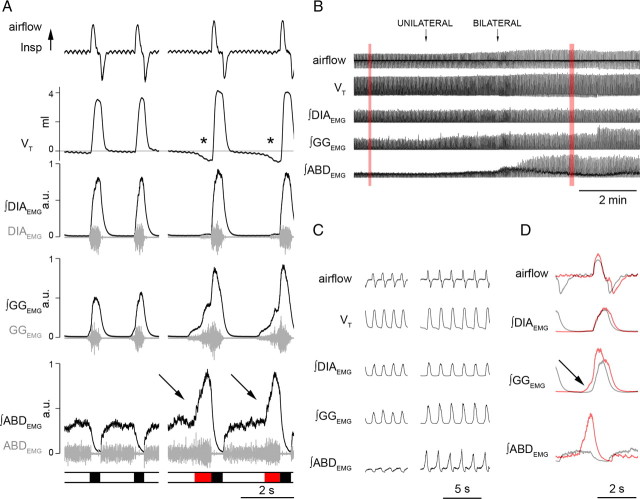
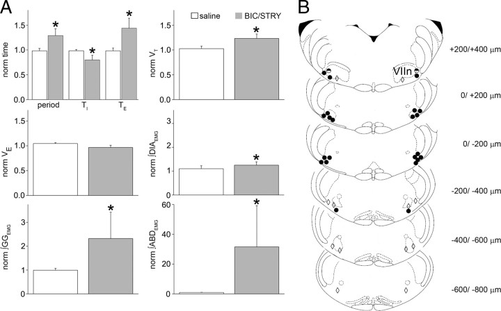
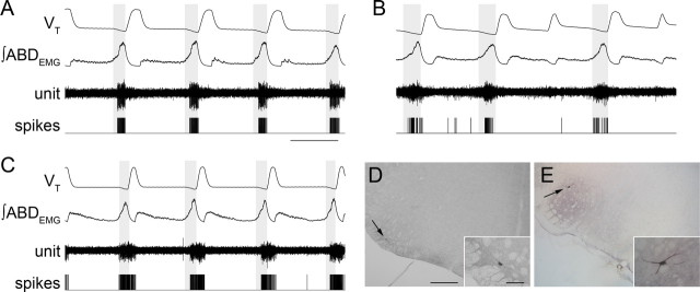
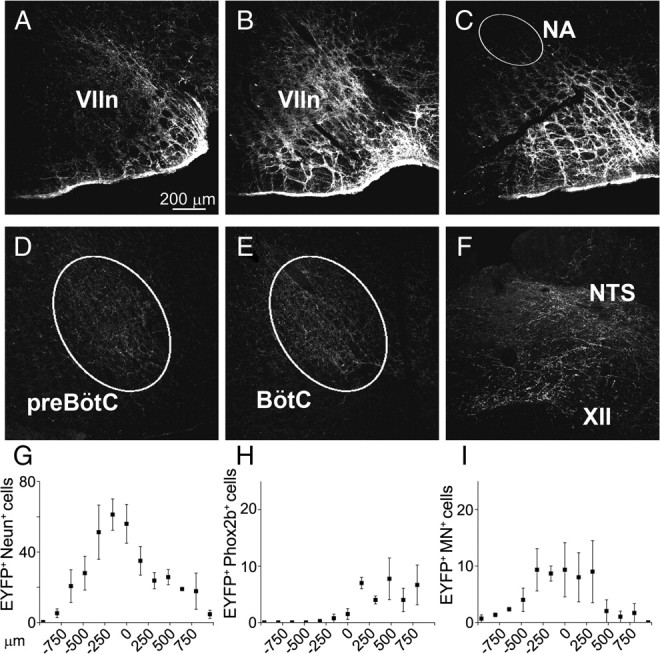
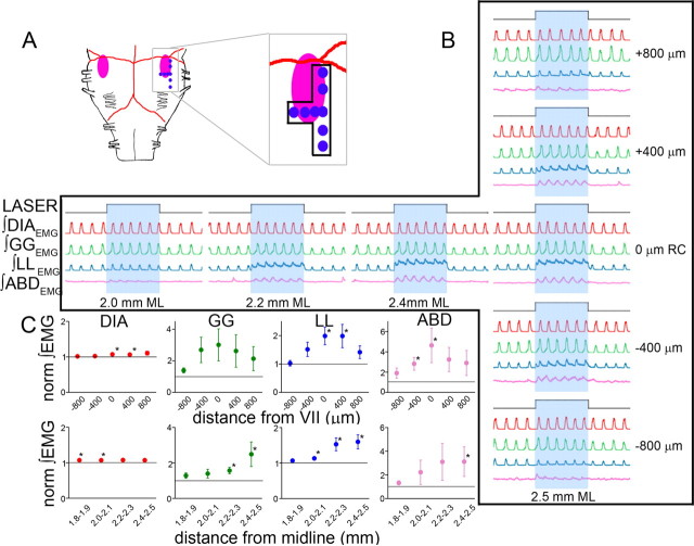
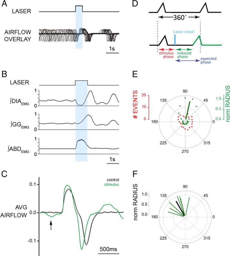
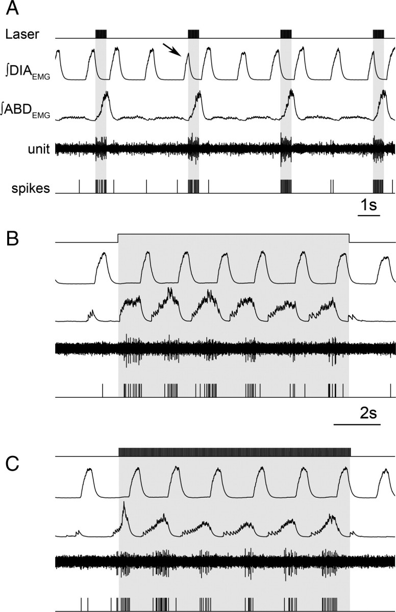
Data from perinatal and juvenile rodents support our hypothesis that the preBötzinger complex generates inspiratory rhythm and the retrotrapezoid nucleus-parafacial respiratory group (RTN/pFRG) generates active expiration (AE). Although the role of the RTN/pFRG in adulthood is disputed, we hypothesized that its rhythmogenicity persists but is typically silenced by synaptic inhibition. We show in adult anesthetized rats that local pharmacological disinhibition or optogenetic excitation of the RTN/pFRG can generate AE and transforms previously silent RTN/pFRG neurons into rhythmically active cells whose firing is correlated with late-phase active expiration. Brief excitatory stimuli also reset the respiratory rhythm, indicating strong coupling of AE to inspiration. The AE network location in adult rats overlaps with the perinatal pFRG and appears lateral to the chemosensitive region of adult RTN. We suggest that (1) the RTN/pFRG contains a conditional oscillator that generates AE, and (2) at rest and in anesthesia, synaptic inhibition of RTN/pFRG suppresses AE.


Figures

Figure 1.
Figure 1.
Local application of BIC/STRY in RTN/pFRG induces long-lasting AE. A, Respiratory airflow, VT, DIAEMG, GGEMG, ABDEMG (gray), and their integrals (black) during passive expiration (left) in respiratory cycles at rest and during AE on stimulation (right). The bar at bottom indicates inspiration (black) and AE (red). During AE, ∫ABDEMG and ∫GGEMG increased and VT dipped below resting levels (asterisks). Integrated traces are plotted from minimum to maximum and scaled [0, 1]. Note the potent increase of ∫ABDEMG at the end of expiratory period during AE. B, Response to unilateral and bilateral application of BIC/STRY (50 μm; 200 nl) in RTN/pFRG. The traces in red boxes are expanded in C. D, Superimposed traces of airflow and EMGs before (black) and after (red) BIC/STRY application. The arrow in D indicates effect of BIC/STRY on GGEMG onset.
Figure 2.
Figure 2.
RTN/pFRG BIC/STRY injections induced AE. A, Respiratory period, TI, TE, VT, V̇E, ∫DIAEMG, ∫GGEMG, ∫ABDEMG calculated from 100 cycles before BIC/STRY injection and after AE onset. Values averaged and normalized in each experiment (n = 4 saline, white; n = 8 BIC/STRY; gray). The asterisk (*) indicates statistical significance (p < 0.05) between preinjection and postinjection. Error bars indicate SEM. B, Representative sections of adult rat brainstem [modified from Paxinos and Watson (1998)]; 0 μm is caudal tip of VIIn. Each symbol represents an injection site. The circles identify locations where AE was induced strongly (filled) or weakly (half-filled). The diamonds identify locations that did not induce AE.
Figure 3.
Figure 3.
RTN/pFRG and VII motor neuronal activity during AE. A–C, Three different neurons that were silent at rest, and rhythmic after induction of AE. Traces (top to bottom), VT, ∫ABDEMG, neuronal activity, and derived spikes. The shaded boxes demark late expiration. Calibration: 2 s. D, E, Photomicrographs of transverse sections of rat brainstem illustrating juxtacellular labeling of late expiratory neuron recorded shown in B (D, arrow) and VII motoneuron (E, arrow). The insets show higher magnification of neurons. Scale bars: D, E, 500 μm; D, E, insets, 100 μm.
Figure 4.
Figure 4.
EYFP expression 4 weeks after SYN-ChR2-EYFP lentivirus injected in rat RTN/pFRG. A–C, EYFP expression in cell bodies and fibers surrounding rostral VIIn, caudal VIIn, and caudal to the VIIn, respectively. D–F, EYFP expression in fibers in preBötC, BötC, hypoglossal nucleus (XII), and NTS (NA, nucleus ambiguus). G–I, Rostrocaudal distribution of EYFP+/NeuN+, EYFP+/Phox2b+, and EYFP+/MN+ double-labeled somata in relationship to caudal pole of the VIIn (n = 4).
Figure 5.
Figure 5.
Photostimulation on ventral medullary surface over RTN/pFRG in SYN-ChR2-EYFP-injected rats induced AE. A, Diagram of ventral brainstem surface (VIIn: pink) indicating photostimulation sites (blue dots correspond to sets of traces in B). B, Effects of 10 s photostimulus (top trace) on DIAEMG (red), GGEMG (green), LLEMG (blue), and ABDEMG (pink). Right, ±400 μm rostrocaudal (RC) intervals; middle, ±200 μm mediolateral (ML) intervals. C, Effect of 10 s photostimulus along the rostrocaudal (top; n = 5) and mediolateral (bottom; n = 4) axes on peak DIAEMG (red), GGEMG (green), LLEMG (blue), and ABDEMG (pink). The horizontal lines represent normalized control values before photostimulation. The asterisks indicate p < 0.05 between control and photostimulation.
Figure 6.
Figure 6.
Brief photostimulation of RTN/pFRG neurons resets respiratory cycle. A, Superposition of respiratory airflow traces (n = 35) during unilateral 500 ms pulse photostimulation, which resets and aligns the subsequent traces. B, Averaged traces for ∫DIAEMG, ∫GGEMG, and ∫ABDEMG show reset of EMG on photostimulation (1 s pulse; n = 35 cycles). Data normalized from 0 to 1. C, Averaged flow before (black) and after (green) 500 ms photostimulation (n = 35 cycles) aligned to inspiratory onset. The arrow indicates expiratory airflow induced by photostimulation. The dotted line indicates the 95% confidence interval for the mean. D, Phases in reset phase analysis. Top trace, Control respiratory cycle (from 0 to 360°, measured from the onset of one inspiration to the next). Bottom trace, Respiratory cycle during stimulation. Stimulus phase, Onset of photostimulation with respect to the phase of respiration; induced phase, onset of inspiration subsequent to delivery of photostimulation; expected phase, expected onset of the next inspiratory cycle with respect to stimulus onset if photostimulation had no effect (360° minus stimulus phase). E, Distribution of events for stimulus and induced phases during multiple trials of 500 ms photostimulation delivered during the same experiment (A). Although stimuli were distributed randomly across all phases of respiration (red dots), the stimulus-induced phase values (green dots) demonstrated clustering at a tight range of preferred phase angles. The average preferred angle for the induced phase is overlaid as a green vector. The left scale bar indicates radial distance for number of events (0–20) for stimulus or induced phase. The right scale bar indicates normalized radial length for vector (0–1, with values close to 1 being indicative of low dispersion of angles and significant phase preferences in polar distributions). F, Distribution of preferred phases of stimulus-induced respiration for seven separate experiments plotted as vectors in normalized length units (green) and the calculated grand average (black) across all experiments.
Figure 7.
Figure 7.
Late expiratory neuron activated by RTN/pFRG photostimulation. A, Brief trains (20 Hz, 10 ms) of photostimulation activate RTN/pFRG neurons and recruits ABDEMG. Note interruption of inspiratory DIAEMG (arrow) on photostimulation with sudden recruitment of ABDEMG and expiratory neuron activity. Continuous (10 s) (B) and brief trains (20 Hz, 10 ms for 10 s) (C) promote activation of ABDEMG bursts and neuron fires only during the late expiratory phase.

Similar articles

Cited by

References

    1. Abbott SB, Stornetta RL, Fortuna MG, Depuy SD, West GH, Harris TE, Guyenet PG. Photostimulation of retrotrapezoid nucleus phox2b-expressing neurons in vivo produces long-lasting activation of breathing in rats. J Neurosci. 2009a;29:5806–5819. - PMC - PubMed
    1. Abbott SB, Stornetta RL, Socolovsky CS, West GH, Guyenet PG. Photostimulation of channelrhodopsin-2 expressing ventrolateral medullary neurons increases sympathetic nerve activity and blood pressure in rats. J Physiol. 2009b;587:5613–5631. - PMC - PubMed
    1. Abdala AP, Rybak IA, Smith JC, Paton JF. Abdominal expiratory activity in the rat brainstem-spinal cord in situ: patterns, origins and implications for respiratory rhythm generation. J Physiol. 2009;587:3539–3559. - PMC - PubMed
    1. Abraham KA, Feingold H, Fuller DD, Jenkins M, Mateika JH, Fregosi RF. Respiratory-related activation of human abdominal muscles during exercise. J Physiol. 2002;541:653–663. - PMC - PubMed
    1. Adamantidis AR, Zhang F, Aravanis AM, Deisseroth K, de Lecea L. Neural substrates of awakening probed with optogenetic control of hypocretin neurons. Nature. 2007;450:420–424. - PMC - PubMed

Publication types