Comparative Study
. 2011 Feb 23;31(8):2916-24.
doi: 10.1523/JNEUROSCI.4771-10.2011.

Conformational flexibility of the ligand-binding domain dimer in kainate receptor gating and desensitization

Affiliations
Comparative Study

Conformational flexibility of the ligand-binding domain dimer in kainate receptor gating and desensitization

Naushaba Nayeem et al. J Neurosci. .

Abstract

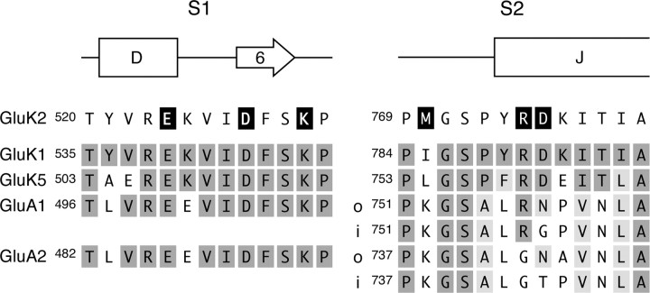
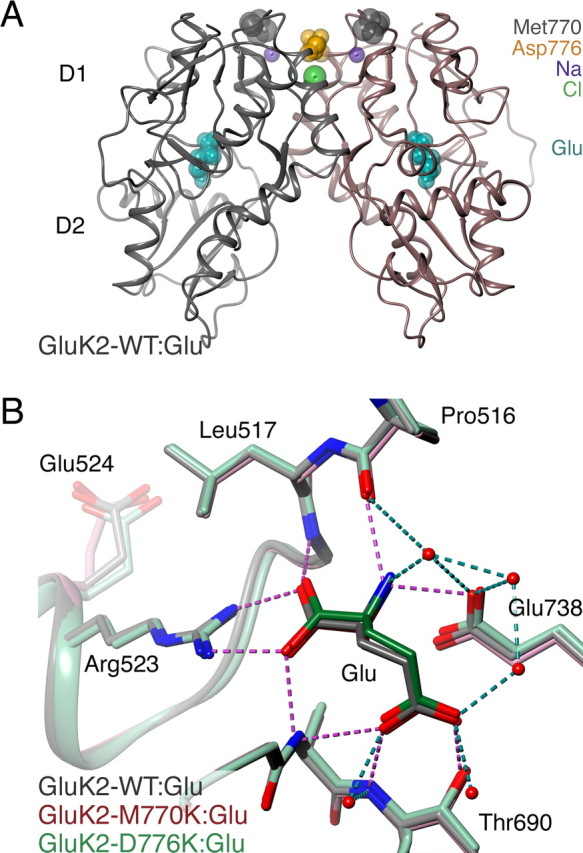
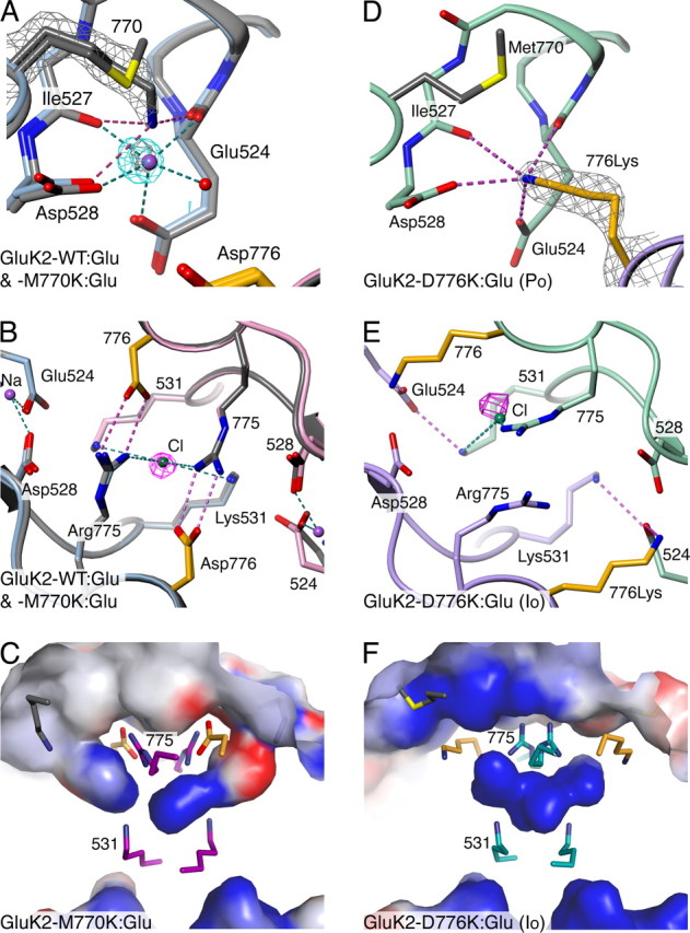
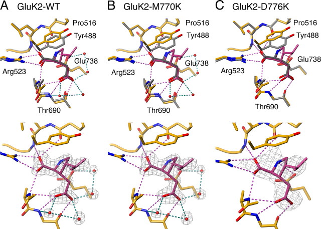
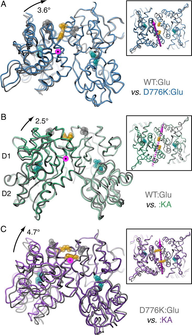
AMPA- and kainate (KA)-selective ionotropic glutamate receptors (iGluRs) respond to agonist by opening (gating), then closing (desensitizing) in quick succession. Gating has been linked to agonist-induced changes within the ligand-binding domain (LBD), and desensitization to rearrangement of a dimer formed by neighboring LBDs. To explore the role of dimer conformation in both gating and desensitization, we compared the conformational effects of two kainate receptor mutants. The first, GluK2-D776K, blocks desensitization of macroscopic current responses ("macroscopic desensitization"). The second, GluK2-M770K, accelerates macroscopic desensitization and eliminates the effects of external ions on channel kinetics. Using structures determined by x-ray crystallography, we found that in both mutants the introduced lysines act as tethered cations, displacing sodium ions from their binding sites within the dimer interface. This results in new inter- and intra-protomer contacts in D776K and M770K respectively, explaining the effects of these mutations on dimer stability and desensitization kinetics. Further, chloride binding was unaffected by the M770K mutation, even though binding of sodium ions has been proposed to promote dimer stability by stabilizing anion binding. This suggests sodium binding may affect receptor function more directly than currently supposed. Notably, we also observed a ligand-specific shift in dimer conformation when comparing LBD dimers in complex with glutamate or the partial agonist KA, revealing a previously unidentified role for dimer orientation in iGluR gating.


Figures

Figure 1.
Figure 1.
Alignment of apical regions of selected kainate- and AMPA-selective receptor subunits. Alignment of S1 and S2 segments around the apical cluster in the GluK2 LBD with equivalent regions in other kainate (GluK1 and GluK5) and AMPA (GluA1 and GluA2) receptor subunits. Residue numbering is for the full-length receptor, except GluA2 where it is for the mature polypeptide. α-Helices (boxes) and a β-strand (arrow) are indicated above the sequence. In S2, sequences of GluA1 and GluA2 are shown for both flop (o) and flip (i) splice variants, as these differ at the sites homologous to Arg775 and Asp776. GluA2 residue 743 is changed by RNA editing from Arg to Gly (the “R/G site”). Key residues mentioned in the text are highlighted for GluK2.
Figure 2.
Figure 2.
LBD dimer overview and ligand-binding site in GluK2-WT:Glu, GluK2–M770K:Glu, and GluK2–D776K:Glu. A, Diagram of GluK2-WT:Glu dimer, showing ligand (cyan), the residues mutated in this study (Met770, gray; Asp776, orange) and ions (sodium, purple; chloride, green) as spheres. The D1 and D2 domains are indicated. B, Ligand interactions at the binding sites of GluK2-WT:Glu (gray), GluK2–M770K:Glu (pink), and GluK2–D776K:Glu (Po form; green) (all protomer B). The structures of GluK2-WT and GluK2–M770K superimpose almost exactly. Side-chains and main-chain sections that interact with the ligand Glu are displayed as sticks. Glu524, which forms part of the cation-binding pocket, is also shown. Interacting waters (from WT only) are shown as nonbonded spheres. Dashed lines represent interactions in GluK2-WT between ligand and either protein (magenta) or waters (blue-green).
Figure 3.
Figure 3.
Comparison of apical interactions in GluK2-WT:Glu, GluK2-M770K:Glu, and GluK2-D776K:Glu. A, Superimposed view of cation-binding site in protomers A of GluK2-WT:Glu (gray) and GluK2-M770K:Glu (blue). Sodium (purple) and an interacting solvent molecule are shown for GluK2-WT only (blue-green dashes show contacts). Only the main conformer of residue 770 is shown for GluK2-M770K (magenta dashes show hydrogen-bonds). Omit-map (gray mesh, |2FobsFcalccalc contoured at 1.3 σ) and difference-Fourier (cyan mesh, contoured at 5 σ) electron density is shown for GluK2-M770K around lysine 770 and the sodium only. The difference-map was calculated for a model lacking sodium and with full occupancy of the main lysine 770 conformer. B, Overlay of GluK2-WT:Glu and GluK2-M770K:Glu viewed from along the dimer twofold. Sodium and chloride (green) ions and atom contacts (magenta and blue-green dashes) are displayed for GluK2-WT only. Anomalous difference-Fourier electron density (magenta mesh, contoured at 4 σ) is displayed for GluK2-M770K. Displacement of the peak relative to the ion position results from slight non-isomorphism between datasets. C, Electrostatic surface potential (red, −15kbT/ec; blue, +15kbT/ec) for GluK2-M770K:Glu, calculated for protein in the absence of bound ions. The dimer interface is viewed from within protomer B. Ion-binding site residues as shown as sticks (residues 770 and 776 colored gray and orange respectively). D, GluK2-D776K:Glu (Po crystal form) displayed as in A. Protomers A (purple) and B (green) are shown, with omit-map electron density for the lysine 776 only (contoured at 1.5 σ). E, F, Views of GluK2-D776K:Glu (Io form), displayed as in B and C, respectively.
Figure 4.
Figure 4.
Kainate binding to GluK2-WT, GluK2-M770K and GluK2-D776K. A, Comparison of ligand-binding sites in Glu and KA complexes (top) and |2FobsFcalccalc electron density maps (bottom) for GluK2-WT. Protomers B are shown. The Glu complex is displayed as gray sticks, and the KA complex in orange (ligand in dark gray and purple respectively). Interactions between kainate and protein are shown as dashed magenta lines, and those involving waters are shown as dashed blue-green lines. In the bottom the electron density map was calculated before kainate was added to the model. Rwork and Rfree (%) for the model at this stage were 25.0 and 27.5. Electron density is shown around the ligand and waters only, and contoured at 1.5 σ. B, C, Equivalent views for GluK2-M770K and GluK2-D776K (Po crystal forms) respectively, displayed as in panel (A). For GluK2-D776K:KA, protomer C is shown. The Rwork and Rfree values for the maps are 26.1 and 28.7 for GluK2-M770K and 23.1 and 27.1 for GluK2-D776K. Maps are contoured at either 1.5 σ (B) or 1.3 σ (C).
Figure 5.
Figure 5.
Hinged and pivoted movements in the LBD dimer interface. A, GluK2-WT:Glu (gray) and GluK2-D776K:Glu (Io crystal form; blue) aligned on the D1 domains of protomers A only (right side), to highlight shifts in the dimer interface. The main panel is viewed along the rotational axis (magenta with black center), and the boxed inset is viewed from above the twofold to show vector direction (dashed magenta arrow). For orientation ligand (cyan) and residues 770 (gray, main panel only) and 776 (orange) are shown as sticks/space-fill. B, C, Equivalent views of GluK2-WT:Glu (gray) and GluK2-WT:KA (green) and GluK2-D776K:Glu (Io crystal form; gray) and GluK2-WT:KA (purple), respectively, aligned and displayed as in A. In all parts the magnitude of the rotational component is indicated at the top left, where its effect is most evident. Movement due to cleft-closure is apparent comparing Glu and KA complexes in B and C (main panels, right-side protomer D2 domains), but not comparing GluK2-WT:Glu and GluK2-WT-D776K:Glu (A).

Similar articles

Cited by

References

    1. Adams PD, Grosse-Kunstleve RW, Hung LW, Ioerger TR, McCoy AJ, Moriarty NW, Read RJ, Sacchettini JC, Sauter NK, Terwilliger TC. PHENIX: building new software for automated crystallographic structure determination. Acta Crystallogr D Biol Crystallogr. 2002;58:1948–1954. - PubMed
    1. Armstrong N, Gouaux E. Mechanisms for activation and antagonism of an AMPA-sensitive glutamate receptor: crystal structures of the GluR2 ligand binding core. Neuron. 2000;28:165–181. - PubMed
    1. Armstrong N, Jasti J, Beich-Frandsen M, Gouaux E. Measurement of conformational changes accompanying desensitization in an ionotropic glutamate receptor. Cell. 2006;127:85–97. - PubMed
    1. Baker NA, Sept D, Joseph S, Holst MJ, McCammon JA. Electrostatics of nanosystems: application to microtubules and the ribosome. Proc Natl Acad Sci U S A. 2001;98:10037–10041. - PMC - PubMed
    1. Birdsey-Benson A, Gill A, Henderson LP, Madden DR. Enhanced efficacy without further cleft closure: reevaluating twist as a source of agonist efficacy in AMPA receptors. J Neurosci. 2010;30:1463–1470. - PMC - PubMed

Publication types

LinkOut - more resources