

Investigation of the neural control of cough and cough suppression in humans using functional brain imaging
- PMID: 21414916
- PMCID: PMC6623770
- DOI: 10.1523/JNEUROSCI.4597-10.2011
Investigation of the neural control of cough and cough suppression in humans using functional brain imaging
Abstract
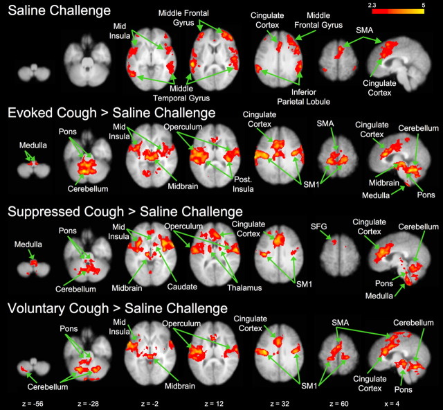
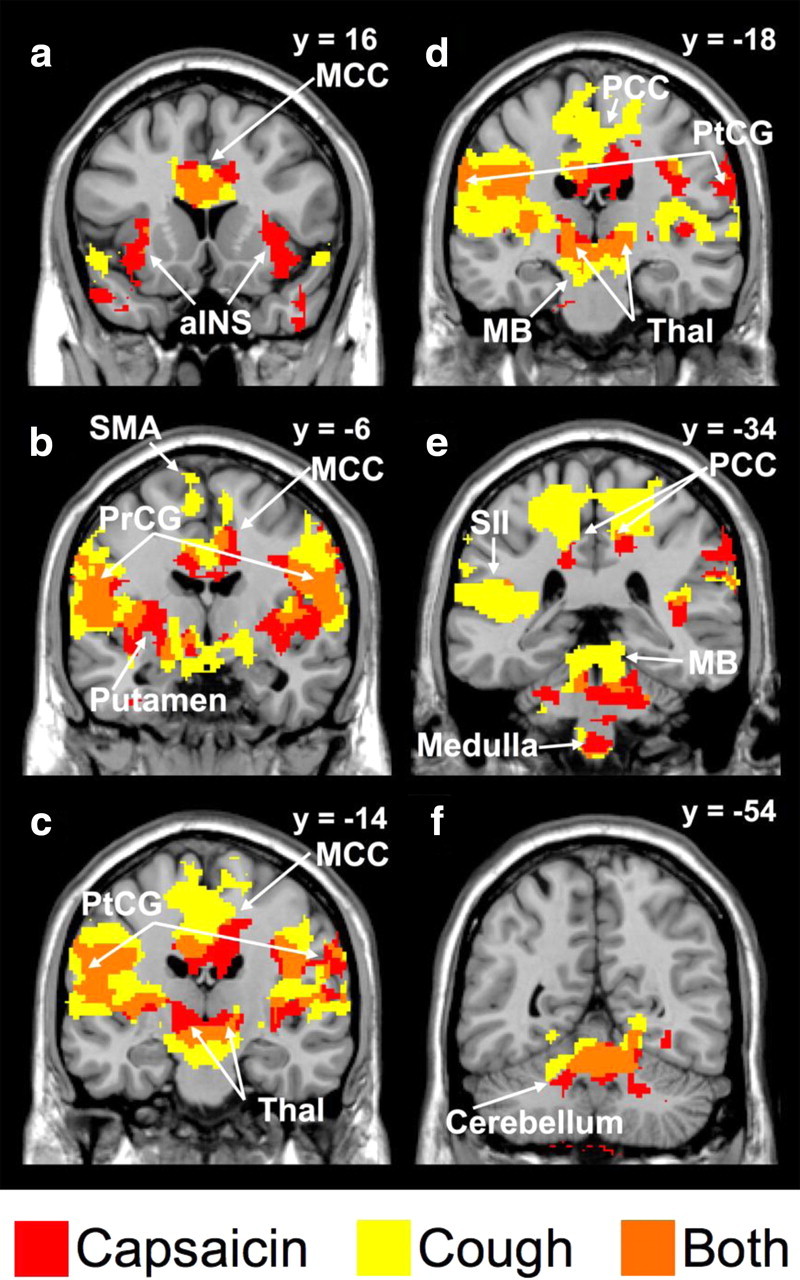
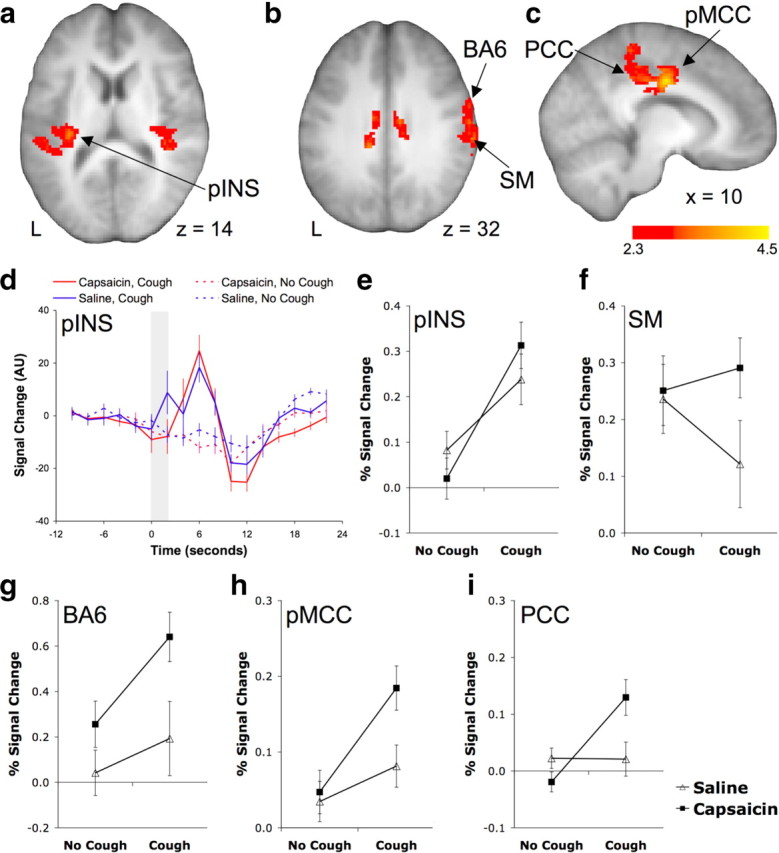
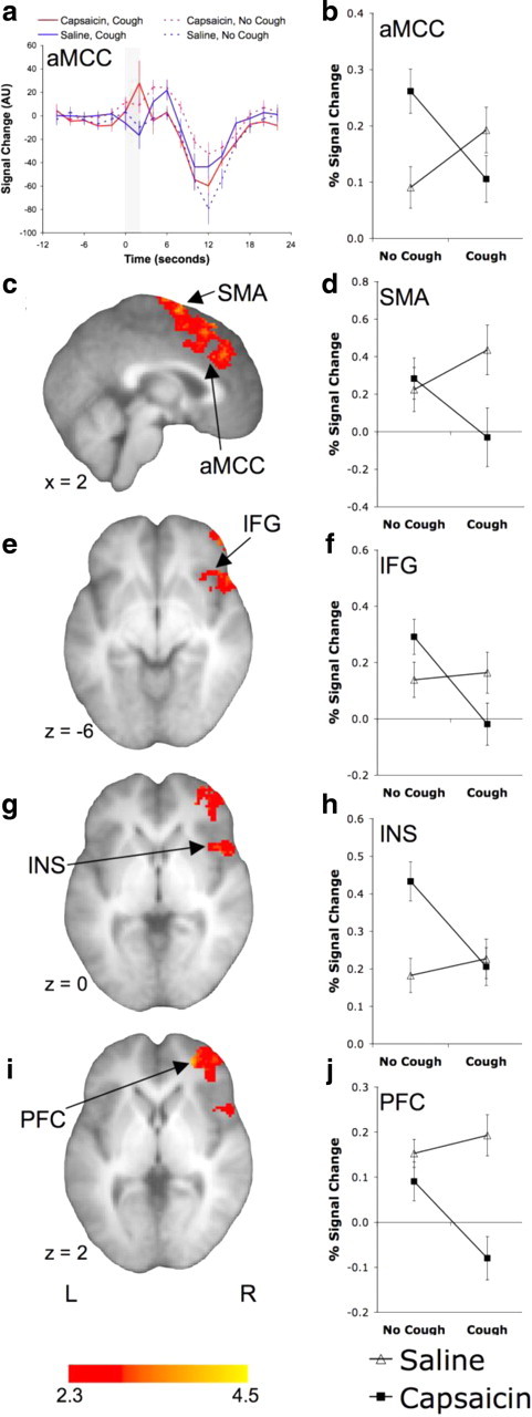
Excessive coughing is one of the most common reasons for seeking medical advice, yet the available therapies for treating cough disorders are inadequate. Humans can voluntarily cough, choose to suppress their cough, and are acutely aware of an irritation that is present in their airways. This indicates a significant level of behavioral and conscious control over the basic cough reflex pathway. However, very little is known about the neural basis for higher brain regulation of coughing. The aim of the present study was to use functional brain imaging in healthy humans to describe the supramedullary control of cough and cough suppression. Our data show that the brain circuitry activated during coughing in response to capsaicin-evoked airways irritation is not simply a function of voluntarily initiated coughing and the perception of airways irritation. Rather, activations in several brain regions, including the posterior insula and posterior cingulate cortex, define the unique attributes of an evoked cough. Furthermore, the active suppression of irritant-evoked coughing is also associated with a unique pattern of brain activity, including an involvement of the anterior insula, anterior mid-cingulate cortex, and inferior frontal gyrus. These data demonstrate for the first time that evoked cough is not solely a brainstem-mediated reflex response to irritation of the airways, but rather requires active facilitation by cortical regions, and is further regulated by distinct higher order inhibitory processes.
Figures
Similar articles
-
Neural correlates of cough hypersensitivity in humans: evidence for central sensitisation and dysfunctional inhibitory control.Thorax. 2016 Apr;71(4):323-9. doi: 10.1136/thoraxjnl-2015-207425. Epub 2016 Feb 9. Thorax. 2016. PMID: 26860344
-
Representation of capsaicin-evoked urge-to-cough in the human brain using functional magnetic resonance imaging.Am J Respir Crit Care Med. 2007 Aug 15;176(4):327-32. doi: 10.1164/rccm.200612-1856OC. Epub 2007 Jun 15. Am J Respir Crit Care Med. 2007. PMID: 17575093
-
Mapping supramedullary pathways involved in cough using functional brain imaging: comparison with pain.Pulm Pharmacol Ther. 2009 Apr;22(2):90-6. doi: 10.1016/j.pupt.2008.08.003. Epub 2008 Aug 28. Pulm Pharmacol Ther. 2009. PMID: 18804546 Review.
-
Brain activity associated with placebo suppression of the urge-to-cough in humans.Am J Respir Crit Care Med. 2013 Nov 1;188(9):1069-75. doi: 10.1164/rccm.201306-1079OC. Am J Respir Crit Care Med. 2013. PMID: 24093551
-
Cough-related neural processing in the brain: a roadmap for cough dysfunction?Neurosci Biobehav Rev. 2014 Nov;47:457-68. doi: 10.1016/j.neubiorev.2014.09.018. Epub 2014 Oct 6. Neurosci Biobehav Rev. 2014. PMID: 25301754 Review.
Cited by
-
Two cortical representations of voice control are differentially involved in speech fluency.Brain Commun. 2021 Jan 5;3(2):fcaa232. doi: 10.1093/braincomms/fcaa232. eCollection 2021. Brain Commun. 2021. PMID: 33959707 Free PMC article.
-
Neurobiology of Coughing in Children.J Clin Med. 2023 Nov 24;12(23):7285. doi: 10.3390/jcm12237285. J Clin Med. 2023. PMID: 38068335 Free PMC article. Review.
-
Sensorimotor circuitry involved in the higher brain control of coughing.Cough. 2013 Mar 6;9(1):7. doi: 10.1186/1745-9974-9-7. Cough. 2013. PMID: 23497672 Free PMC article.
-
Neural Mechanisms Underlying the Coughing Reflex.Neurosci Bull. 2023 Dec;39(12):1823-1839. doi: 10.1007/s12264-023-01104-y. Epub 2023 Aug 22. Neurosci Bull. 2023. PMID: 37606821 Free PMC article. Review.
-
Anatomy and neurophysiology of cough: CHEST Guideline and Expert Panel report.Chest. 2014 Dec;146(6):1633-1648. doi: 10.1378/chest.14-1481. Chest. 2014. PMID: 25188530 Free PMC article. Review.
References
-
- Andersson JL. How to estimate global activity independent of changes in local activity. Neuroimage. 1997;6:237–244. - PubMed
Publication types
MeSH terms
LinkOut - more resources
Full Text Sources
Medical