. 2011 Jul 1;17(13):4378-88.
doi: 10.1158/1078-0432.CCR-10-2285. Epub 2011 Mar 17.

Dual mTORC2/mTORC1 targeting results in potent suppressive effects on acute myeloid leukemia (AML) progenitors

Affiliations

Dual mTORC2/mTORC1 targeting results in potent suppressive effects on acute myeloid leukemia (AML) progenitors

Jessica K Altman et al. Clin Cancer Res. .

Abstract

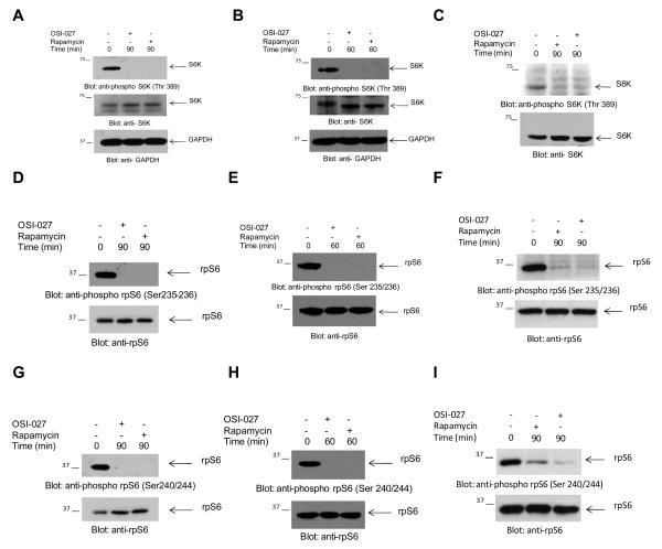
Purpose: To determine whether mTORC2 and rapamycin-insensitive (RI)-mTORC1 complexes are present in acute myeloid leukemia (AML) cells and to examine the effects of dual mTORC2/mTORC1 inhibition on primitive AML leukemic progenitors.

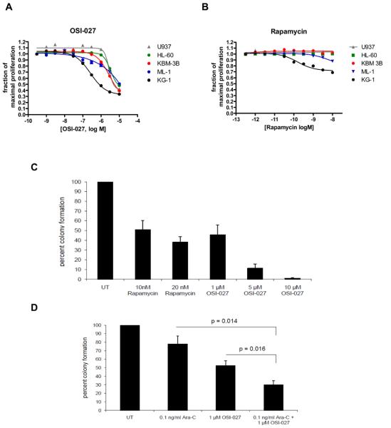
Experimental design: Combinations of different experimental approaches were used, including immunoblotting to detect phosphorylated/activated forms of elements of the mTOR pathway in leukemic cell lines and primary AML blasts; cell-proliferation assays; direct assessment of mRNA translation in polysomal fractions of leukemic cells; and clonogenic assays in methylcellulose to evaluate leukemic progenitor-colony formation.

Results: mTORC2 complexes are active in AML cells and play critical roles in leukemogenesis. RI-mTORC1 complexes are also formed and regulate the activity of the translational repressor 4E-BP1 in AML cells. OSI-027 blocks mTORC1 and mTORC2 activities and suppresses mRNA translation of cyclin D1 and other genes that mediate proliferative responses in AML cells. Moreover, OSI-027 acts as a potent suppressor of primitive leukemic precursors from AML patients and is much more effective than rapamycin in eliciting antileukemic effects in vitro.

Conclusions: Dual targeting of mTORC2 and mTORC1 results in potent suppressive effects on primitive leukemic progenitors from AML patients. Inhibition of the mTOR catalytic site with OSI-027 results in suppression of both mTORC2 and RI-mTORC1 complexes and elicits much more potent antileukemic responses than selective mTORC1 targeting with rapamycin.


Figures

Figure 1
Figure 1
Presence of mTORC2 and mTORC1 complexes in AML cells and differential regulation by rapamycin or OSI-027. A. U937 cells were incubated with or without OSI-027 (10 μM) or rapamycin (20nM) for 90 minutes. Cell lysates were resolved by SDS-PAGE and immunoblotted with an antibody against the phosphorylated form of AKT on serine 473. Subsequently, the same blot was re-probed with antibodies against AKT or GAPDH, to control for equal protein loading. B. Primary leukemic blasts from a patient with AML were incubated with or without OSI-027 (10 μM) or rapamycin (20 nM) for 90 minutes. Cell lysates were resolved by SDS-PAGE and immunoblotted with an antibody against the phosphorylated form of AKT on serine 473. Subsequently, the same blot was re-probed with antibodies against AKT or GAPDH, to control for protein loading. C-D. U937 cells were incubated with or without OSI-027 (5 μM) or rapamycin (20 nM) for 90 minutes. Cell lysates were resolved by SDS-PAGE and immunoblotted with antibodies against the phosphorylated form mTOR on serine 2448 (C) or the phosphorylated form of mTOR on serine 2481 (D). The blots were then re-probed with an antibody against mTOR, to control for protein loading. E. KG1 cells were incubated with or without OSI-027 (10 μM) or rapamycin (20nM) for 24 hours and cell lysates were resolved and immunoblotted with an antibody against the phosphorylated form of mTOR on serine 2481. The same blot was subsequently re-probed with an antibody against mTOR. F. Leukemic blasts from a patient with AML were incubated with or without OSI-027 (10 μM) or rapamycin (20nM) for 90 minutes. Cell lysates were resolved by SDS-PAGE and then immunoblotted with an antibody against the phosphorylated form of mTOR on serine 2481. The blot was then re-probed with an antibody against mTOR.
Figure 2
Figure 2
Phosphorylation/activation of S6K and S6 ribosomal protein in AML cells. A-C. U937 cells (A), KG1 cells (B), or primary leukemic blasts from an AML patient (C) were incubated with OSI-027 (10 μM) or rapamycin (20nM) for the indicated times. Cell lysates were resolved by SDS-PAGE and immunoblotted with an antibody against the phosphorylated form of S6K on Thr 389, or with antibodies against S6K or GAPDH, as indicated. D-I. U937 cells (D, G) KG1 cells (E, H), or primary AML blasts (F, I) were incubated in the absence or presence of rapamycin (20nM) or OSI-027 (10 μM) as indicated. Cell lysates were resolved by SDS-PAGE and immunoblotted with an antibody against the phosphorylated form of rpS6 on Ser235/236 (D, E, F) or against the phosphorylated form of rpS6 on Ser240/244 (G, H, I) or against rpS6, as indicated.
Figure 3
Figure 3
Effects of mTOR inhibition on the expression of the S6K-dependent PDCD4 tumor suppressor in AML cells and functional consequences of S6K knockdown. A-B. U937 (A) or KG1 (B) cells were incubated in the absence (lane 1) or presence of OSI-027 at either 5 μM (lane 2) or 10 μM (lane 3) or rapamycin (20 nM) (lane 4) for 24 hours. Cell lysates were resolved by SDS-PAGE and immunoblotted with an antibody against PDCD4. The same blots were re-probed with an antibody against tubulin, to control for protein loading. C. Cell lysates from lentivirus transduced control-shRNA or S6K1-shRNA U937cells were resolved by SDS-PAGE and immunoblotted with an anti-S6K1 antibody. The same blot was then re-probed with an anti-GAPDH antibody to control for protein loading. D. Leukemic CFU-L colony formation derived from control-shRNA and S6K1-shRNA U937 cells is shown. Data are expressed as percentages of colony formation of untreated control-shRNA transduced cells and represent means ± S.E. of 5 independent experiments.
Figure 4
Figure 4
OSI-027, but not rapamycin, inhibits phosphorylation of the 4E-BP1 repressor of mRNA translation and blocks formation of complexes required for cap-dependent mRNA translation. A-B. U937 or KG1 cells were incubated in the absence or presence of OSI-027 (10 μM) or rapamycin (20nM) for 90 minutes. Cell lysates were resolved by SDS-PAGE and immunoblotted with an antibody against the phosphorylated form of 4E-BP1 on threonine 37/46 (A) or serine 65 (B). Subsequently, the same blots were re-probed with an antibody against 4E-BP1 or against GAPDH, to control for protein loading. C. KG1 cells were incubated in the absence or presence of OSI-027 (10 μM) or rapamycin (20nM) for 60 minutes. Cell lysates were resolved by SDS-PAGE and immunoblotted with an antibody against 4E-BP1 on threonine 70. The same blot was re-probed with an antibody against 4E-BP1 or against GAPDH. D. U937 cells were treated with increasing concentrations of rapamycin or OSI, as indicated for 90 minutes. Cell lysates were resolved by SDS-PAGE and immunoblotted with antibodies against the phosphorylated form of rpS6 at serine 240/244, the phosphorylated form of 4E-BP1 on threonine 37/46, or against GAPDH as indicated. E-F. Leukemic blasts from a patient with AML were incubated in the absence or presence of OSI-027 (10 μM) or rapamycin (20nM) for 90 minutes. Cell lysates were resolved by SDS-PAGE and immunoblotted with an antibody against 4E-BP1 on threonine 37/46 (E) or serine 65 (F). The same blot was re-probed with an antibody against 4E-BP1 and then GAPDH, to assess for equivalent protein loading. G. U937 cells were treated with OSI-027 (10 μM) or rapamycin (20nM) for 90 minutes. Cell lysates were immunoprecipitated with an anti-eIF4E antibody and immunoprecipitates were immunoblotted with antibodies against 4E-BP1, eIF4A, eIF4G or eIF4E, as indicated. A signal seen above the eIF4G band in the anti-eIF4G immunoblot in the OSI-027-treatment lane is an artifact and does not represent a slower migrating form of eIF4G.
Figure 5
Figure 5
Effects of OSI-027 on polysomal assembly and mRNA translation of the cyclin D1 gene in AML cells. A. U937 cells were treated with solvent control (DMSO), rapamycin (20nM) or OSI-027 (10 μM) for 24 hours and the lysates were layered on a 10-50% sucrose gradient. The gradients were subjected to ultracentrifugation and fractions were collected by continuous monitoring of OD at 254nm. The OD 254nm is shown as a function of gradient depth for each treatment and the polysomal peaks are indicated. B. The area under the polysome peaks and polysome (PS) + monosome (MS) peaks was quantified for each treatment using Image J software. The ratio of area under the polysomal and polysomal plus monosomal peaks was calculated for each treatment and is represented as percent control (DMSO). The data represent means + SE from 2 independent experiments. C. The polysomal fractions were pooled and the total RNA was isolated. Quantitative real time RT-PCR assay to determine the Cyclin D1 mRNA expression in polysomal fractions was carried out using HPRT as control. Data are expressed as fold change as compared to DMSO treated samples and represent means + SE of 2 independent experiments. D. U937 cells were incubated with or without OSI-027 (1 μM) for the indicated times. Cell lysates were resolved by SDS-PAGE and immunoblotted with an antibody against cyclin D1. Subsequently, the same blot was re-probed with antibodies against GAPDH, to control for equal protein loading.
Figure 6
Figure 6
Inhibitory effects of OSI-027 on leukemic cell lines and primary leukemic progenitor colony formation from AML patients. A-B. The sensitivity of 5 leukemic cell lines to growth inhibition by OSI-027 (A) or rapamycin (B) is shown. Effects of varying concentrations of each drug at 72 hours are expressed as dose-response curves, and error bars, where visible, show the standard error of the mean for three replicates per data point. Results shown are typical of three or more independent experiments. C. Peripheral blood mononuclear cells from 5 patients with AML were plated in methylcellulose culture assay system with increasing concentrations of rapamycin or OSI-027, as indicated. Data are expressed as percent control of CFU-L colony numbers for untreated cells. Means +/− S.E. of the values from 5 experiments using different patient samples are shown. Paired t test analysis, comparing the effects of OSI–027 (5 μM) to the effects of rapamycin (20 nM), showed p value = 0.0034. D. Peripheral blood mononuclear cells from three patients with AML were plated in methylcellulose culture assay system with Ara-C (0.1 ng/ml) and/or OSI-027 (1 μM), as indicated. Data are expressed as percent control of leukemic colonies for untreated cells and represent means +/− S.E. of 3 experiments. Paired t test analysis of the combination of Ara-C plus OSI-027 compared with Ara-C alone, showed a p= 0.014 and as compared with OSI-027 alone, p= 0.016.

Similar articles

Cited by

References

    1. Lowenberg B, Downing JR, Burnett A. Acute myeloid leukemia. N Engl J Med. 1999;341:1051–62. - PubMed
    1. Tallman MS, Gilliland DG, Rowe JM. Drug therapy for acute myeloid leukemia. Blood. 2005;106:1154–63. - PubMed
    1. Gilliland DG, Tallman MS. Focus on acute leukemias. Cancer Cell. 2002;1:417–20. - PubMed
    1. Cammenga J. Gatekeeper pathways and cellular background in the pathogenesis and therapy of AML. Leukemia. 2005;19:1719–28. - PubMed
    1. Fathi AT, Grant S, Karp JE. Exploiting cellular pathways to develop new treatment strategies for AML. Cancer Treat Rev. 2010;36:142–50. - PMC - PubMed

Publication types

MeSH terms