

Rapid flow assessment of congenital heart disease with high-spatiotemporal-resolution gated spiral phase-contrast MR imaging
- PMID: 21415248
- PMCID: PMC3121014
- DOI: 10.1148/radiol.11101844
Rapid flow assessment of congenital heart disease with high-spatiotemporal-resolution gated spiral phase-contrast MR imaging
Abstract
Purpose: To validate a prospectively triggered spiral phase-contrast magnetic resonance (MR) sequence accelerated with sensitivity encoding (SENSE) in a population of children and adults with congenital heart disease.
Materials and methods: The local research ethics committee approved this study, and written consent was obtained from all patients or guardians. Stroke volumes were quantified in 40 patients (mean age ± standard deviation: 21.4 years ± 13.8, age range: 3.0-61.3 years; 22 male patients aged 3.0-38.0 years [mean age, 17.2 years ± 10.5], 18 female patients aged 4.7-61.3 years [mean age, 26.6 years ± 15.9]) with congenital heart disease in the aorta (n = 40), main pulmonary artery (n = 38), right pulmonary artery (n = 22), and left pulmonary artery (n = 24). Stroke volumes were obtained with (a) breath-hold spiral phase-contrast MR imaging with SENSE, (b) conventional breath-hold cartesian phase-contrast MR imaging, and (c) reference free-breathing phase-contrast MR imaging. Stroke volumes were compared by using repeated-measures analysis of variance, Bland-Altman analysis, and correlation coefficients.
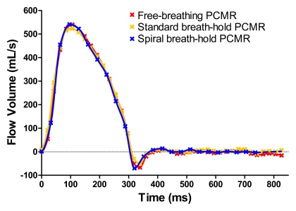
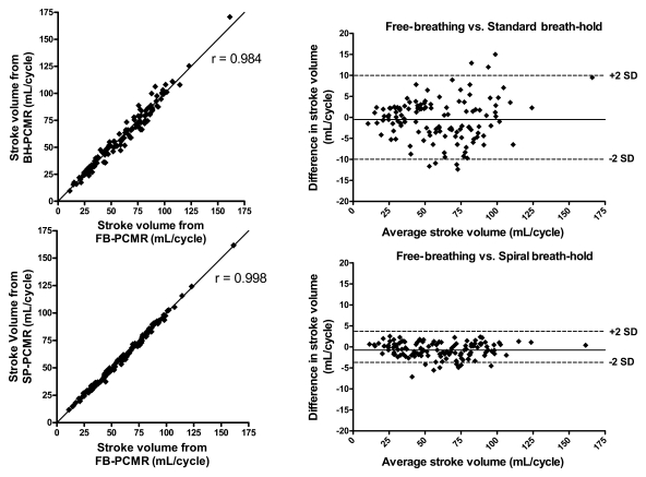
Results: Imaging time with the breath-hold spiral phase-contrast MR sequence was significantly lower than that with the conventional breath-hold phase-contrast MR sequence (~5 seconds vs ~16 seconds, respectively; P < .0001). There was excellent agreement in stroke volumes in all vessels between the reference free-breathing sequence (mean volume, 60.3 mL ± 27.3) and the two breath-hold sequences-spiral SENSE phase-contrast MR imaging (mean volume, 59.5 mL ± 27.1; P < .001) and conventional cartesian phase-contrast MR imaging (mean volume, 59.8 mL ± 27.6; P = .268). The limits of agreement were smaller with the spiral breath-hold sequence than with the conventional breath-hold sequence (-4.4 mL, 2.9 mL vs -10.3 mL, 9.3 mL, respectively); correlation was similar (r = 0.998 vs r = 0.984, respectively).
Conclusion: Flow volumes can be accurately and reliably quantified by using a spiral SENSE phase-contrast MR sequence, with high spatiotemporal resolution obtained in a short breath hold.
Figures
References
-
- Firmin DN, Nayler GL, Klipstein RH, Underwood SR, Rees RSO, Longmore DB. In vivo validation of MR velocity imaging. J Comput Assist Tomogr 1987;11(5):751–756 - PubMed
-
- Beerbaum P, Körperich H, Barth P, Esdorn H, Gieseke J, Meyer H. Noninvasive quantification of left-to-right shunt in pediatric patients: phase-contrast cine magnetic resonance imaging compared with invasive oximetry. Circulation 2001;103(20):2476–2482 - PubMed
-
- Van Rossum AC, Sprenger M, Visser FC, Peels KH, Valk J, Roos JP. An in vivo validation of quantitative blood flow imaging in arteries and veins using magnetic resonance phase-shift techniques. Eur Heart J 1991;12(2):117–126 - PubMed
-
- Rees S, Firmin D, Mohiaddin R, Underwood R, Longmore D. Application of flow measurements by magnetic resonance velocity mapping to congenital heart disease. Am J Cardiol 1989;64(14):953–956 - PubMed
-
- Sakuma H, Kawada N, Kubo H, et al. Effect of breath holding on blood flow measurement using fast velocity encoded cine MRI. Magn Reson Med 2001;45(2):346–348 - PubMed
Publication types
MeSH terms
Grants and funding
LinkOut - more resources
Full Text Sources
Medical
