. 2011 Mar 18;331(6023):1439-43.
doi: 10.1126/science.1200113.

Cardiac myosin activation: a potential therapeutic approach for systolic heart failure

Affiliations

Cardiac myosin activation: a potential therapeutic approach for systolic heart failure

Fady I Malik et al. Science. .

Abstract

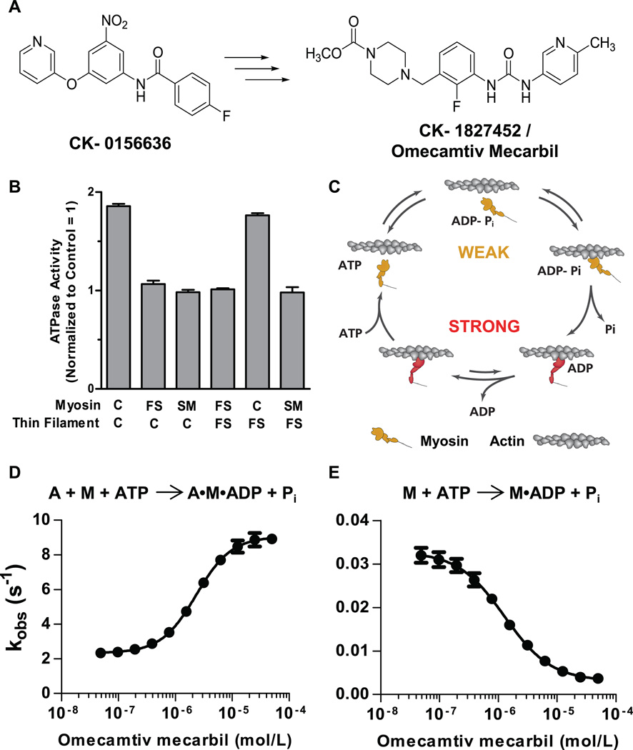
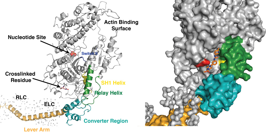
Decreased cardiac contractility is a central feature of systolic heart failure. Existing drugs increase cardiac contractility indirectly through signaling cascades but are limited by their mechanism-related adverse effects. To avoid these limitations, we previously developed omecamtiv mecarbil, a small-molecule, direct activator of cardiac myosin. Here, we show that it binds to the myosin catalytic domain and operates by an allosteric mechanism to increase the transition rate of myosin into the strongly actin-bound force-generating state. Paradoxically, it inhibits adenosine 5'-triphosphate turnover in the absence of actin, which suggests that it stabilizes an actin-bound conformation of myosin. In animal models, omecamtiv mecarbil increases cardiac function by increasing the duration of ejection without changing the rates of contraction. Cardiac myosin activation may provide a new therapeutic approach for systolic heart failure.


Figures

Fig. 1
Fig. 1
Identification and characterization of omecamtiv mecarbil. (A) The chemical structure of the original hit, CK-0156636, and the chemical structure of omecamtiv mecarbil whose optimization and synthesis have been previously reported (7). (B) Target identification of omecamtiv mecarbil using heterologous reconstituted combinations of the troponin-tropomyosin regulated actin-myosin system. Means ± SD are shown for three to five replicates at each condition. ATPase measurements were made at 25°C in 12 mM Pipes, 2 mM MgCl2, 1 mM dithiothreitol at pH 6.8 and pCa 6.75 by using a coupled enzyme system consisting of pyruvate kinase and lactate dehydrogenase and monitoring the oxidation of NADH at 340 nM (14). C, FS, and SM denote cardiac, fast skeletal, and smooth muscle myosin isoforms. Similarly, C and FS denote reconstituted cardiac and fast skeletal thin filaments, respectively. (C) The mechanochemical cycle of myosin. Yellow indicates myosin weakly bound to actin and red indicates myosin strongly bound to actin (adapted from an illustration, courtesy of J. Spudich, Stanford University). The effect of omecamtiv mecarbil on the phosphate release rate is shown for actin (A) plus myosin (M) in panel (D) and of myosin alone in panel (E). Transient-state kinetic measurements of phosphate release from cardiac myosin S1 (n = 3 for each data point, means ± SD) were carried out by rapidly mixing a solution of myosin and ATP with 7-diethylamino-3-({[(2-maleimidyl)ethyl]amino}carbonyl) coumarin–modified phosphate-binding protein (MDCC-PBP) plus or minus actin with a stopped-flow apparatus under single-turnover conditions at 25°C. Phosphate released from myosin binds rapidly to MDCC-PBP, which leads to an increase in its fluorescence (13). The rate of phosphate release is measured at various concentrations of omecamtiv mecarbil to construct a dose response.
Fig. 2
Fig. 2
The proposed binding site for omecamtiv mecarbil to cardiac myosin S1. The ribbon diagram to the left shows the major features of the myosin S1 head. A space-filling model of the myosin structure showing the position of the identified peptide is shown to the right (the structure of the chicken skeletal S1 fragment, PDB ID 2MYS, was used as a model because the cardiac S1 structure has not been determined). The compound was manually fit into the cleft containing the identified labeled peptide. The red residue indicates the labeled amino acid serine 148.
Fig. 3
Fig. 3
The effects of omecamtiv mecarbil in cardiac myocytes. (A) Representative tracings showing that omecamtiv mecarbil (200 nM) increases cardiac myocyte contractility without changing the Ca2+ transient. In contrast, the β-adrenergic agonist isoproterenol (2 nM) increases contractility by increasing the Ca2+ transient. (B) Treatment of myocytes (n = eight myocytes per condition) with the β-adrenergic blocker, carvedilol, at a concentration (200 nM) sufficient to completely block the effect of isoproterenol (20 nM), does not alter the effect of omecamtiv mecarbil (200 nM) on myocyte contractility (FS). Data are means ± SEM.
Fig. 4
Fig. 4
The effects of omecamtiv mecarbil in models of cardiac function. (A) Fractional shortening (FS) of the heart was measured by echocardiography (parasternal long-axis view) with coincident determination of omecamtiv mecarbil plasma concentration in isoflurane anesthetized Sprague-Dawley rats and beagle dogs during 30- to 60-min infusions of drug or vehicle. Placebo-corrected absolute percentage point increases of FS from baseline are plotted against the mean plasma concentration at each infusion dose. Data are means ± SEM, n = five or six rats or four dogs at each dose. (B) Cardiac function in conscious mongrel (NL) and in heart failure (HF) dogs chronically instrumented to measure LV pressure, wall thickness, dimensions, and cardiac output. In NL (n = 5) and HF (n = 5) dogs, omecamtiv mecarbil was administered as a bolus at 0.5 mg/kg of body weight, followed by infusion at 0.5 mg/kg of body weight per hour. Measurements plotted are mean ± SEM values 15 min after the start of drug administration. WT, wall thickening; FS, fractional shortening; SET, systolic ejection time; dP/dt, rate of pressure change; HR, heart rate; SV, stroke volume; CO, cardiac output. (C) Time-dependent elastance is plotted for dobutamine (10 µg/kg per min) and omecamtiv mecarbil (10 min following a 1 mg/kg bolus). The response of each dog is normalized to the magnitude and time of peak elastance at baseline to allow for comparison of the shape of the response. Statistics were performed using a one-way analysis of variance (ANOVA) with a post hoc Student-Newman-Keuls test.

Comment in

References

    1. Lloyd-Jones D, et al. Executive summary: Heart disease and stroke statistics—2010 update: A report from the American Heart Association. Circulation. 2010;121:948. - PubMed
    1. Loehr LR, Rosamond WD, Chang PP, Folsom AR, Chambless LE. Heart failure incidence and survival (from the Atherosclerosis Risk in Communities study) Am. J. Cardiol. 2008;101:1016. - PubMed
    1. Adams KF, Jr, et al. Characteristics and outcomes of patients hospitalized for heart failure in the United States: Rationale, design, and preliminary observations from the first 100,000 cases in the Acute Decompensated Heart Failure National Registry (ADHERE) Am. Heart J. 2005;149:209. - PubMed
    1. McMurray JJV. Clinical practice. Systolic heart failure. N. Engl. J. Med. 2010;362:228. - PubMed
    1. Kass DA, Solaro RJ. Mechanisms and use of calcium-sensitizing agents in the failing heart. Circulation. 2006;113:305. - PubMed

Publication types

MeSH terms