. 2011 Jun;31(6):1300-8.
doi: 10.1161/ATVBAHA.111.223701. Epub 2011 Mar 17.

SDF-1α induction in mature smooth muscle cells by inactivation of PTEN is a critical mediator of exacerbated injury-induced neointima formation

Affiliations

SDF-1α induction in mature smooth muscle cells by inactivation of PTEN is a critical mediator of exacerbated injury-induced neointima formation

Raphael A Nemenoff et al. Arterioscler Thromb Vasc Biol. 2011 Jun.

Abstract

Objective: PTEN inactivation selectively in smooth muscle cells (SMC) initiates multiple downstream events driving neointima formation, including SMC cytokine/chemokine production, in particular stromal cell-derived factor-1α (SDF-1α). We investigated the effects of SDF-1α on resident SMC and bone marrow-derived cells and in mediating neointima formation.

Methods and results: Inducible, SMC-specific PTEN knockout mice (PTEN iKO) were bred to floxed-stop ROSA26-β-galactosidase (βGal) mice to fate-map mature SMC in response to injury; mice received wild-type green fluorescent protein-labeled bone marrow to track recruitment. Following wire-induced femoral artery injury, βGal(+) SMC accumulated in the intima and adventitia. Compared with wild-type, PTEN iKO mice exhibited massive neointima formation, increased replicating intimal and medial βGal(+)SMC, and enhanced vascular recruitment of bone marrow cells following injury. Inhibiting SDF-1α blocked these events and reversed enhanced neointima formation observed in PTEN iKO mice. Most recruited green fluorescent protein(+) cells stained positive for macrophage markers but not SMC markers. SMC-macrophage interactions resulted in a persistent SMC inflammatory phenotype that was dependent on SMC PTEN and SDF-1α expression.

Conclusion: Resident SMC play a multifaceted role in neointima formation by contributing the majority of neointimal cells, regulating recruitment of inflammatory cells, and contributing to adventitial remodeling. The SMC PTEN-SDF-1α axis is a critical regulator of these events.


Figures
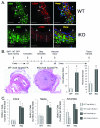
Figure 1
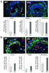
Figure 1. Highly Differentiated SMC Contribute to Injury-Induced Vessel Remodeling and Enhanced Neointima Formation in PTEN iKO Mice
(A) Immunofluorescence staining for βGal and α-SMA on 7-d injured femoral arteries from WT and PTEN iKO mice. Arrows = representative intimal βGal(+);α-SMA(+) SMC; arrowheads = representative adventitial βGal(+) SMC; open arrowheads = representative intimal βGal(+);α-SMA(−) SMC; lines delineate the arterial media. (B) (Top) Time course of experimental protocol. H&E staining of representative arterial lesions in WT and PTEN iKO mice. Medial and intimal areas were measured using SPOT software. Intima-to-media ratios (left) and percent stenotic areas (right) are presented in the graphs. (C) Total numbers of BrdU(+) compared to βGal(+);BrdU(+) SMC in the arterial intima, media, and adventitia at 3-w post-injury were counted separately and the data reported in the graphs. *Different from WT; n=8.
Figure 2
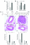
Figure 2. Bone Marrow-Derived Cells are Recruited to Injured Vessels, but do not Differentiate into SMC
(A) Immunofluorescence staining for GFP and α-SMA on 3-w injured femoral arteries from WT and PTEN iKO mice. N=Neointima; M=Media; A=Adventitia. (B) Immunofluorescence staining for βGal and GFP on an uninjured femoral artery. Right panel shows merged image. Arrows = GFP(+);βGal(−) cell in the arterial media; arrowheads = internal elastic lamina. (C) Immunofluorescence staining for GFP and Mac3 on 3-w injured femoral arteries from WT (top panels) and PTEN iKO (bottom panels) mice. Right panels show merged images. Scale bars A&C = 20 μm.
Figure 3
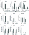
Figure 3. Increased Accumulation of Bone Marrow-Derived Macrophages in Injured Vessels from PTEN iKO Mice
(A) Immunofluorescence staining for GFP on 3-w injured femoral arteries from WT and PTEN iKO mice. Total numbers of GFP(+) cells in the arterial intima, media, and adventitia were counted separately and the data reported in the graphs. (B) Immunofluorescence staining for GFP and BrdU on 3-w injured femoral arteries from WT and PTEN iKO mice. GFP(+);BrdU(+) cells in the arterial intima, media, and adventitia were counted separately and the data reported in the graphs. Arrows = representative GFP(+);BrdU(+) cells; arrowheads = representative GFP(−);BrdU(+) cells. *Different from WT; n=8. N=Neointima; M=Media; A=Adventitia.
Figure 4
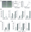
Figure 4. Enhanced Neointima Formation in PTEN iKO mice is Dependent on SDF-1α
(A) qPCR analysis for SDF-1α mRNA in injured femoral arteries from WT and PTEN iKO mice. β-Actin was used for normalization of cDNA. (B) Serum from 7-d and 3-w post-injured WT and PTEN iKO mice was analyzed by ELISA for SDF-1α levels. (C) (Top) Timeline of experimental protocol. H&E staining on 3-w injured femoral arteries from WT and PTEN iKO mice treated with control or SDF-1α neutralizing antibodies. Intima-to-media ratios (left) and percent stenotic areas (right) were calculated and are presented in the graphs. *Different from WT; **PTEN iKO + αSDF-1α different from PTEN iKO + control IgG; n=6.
Figure 5
Figure 5. Increased SMC Proliferation and Accumulation of Bone Marrow-Derived Macrophages in PTEN iKO mice is Dependent on SDF-1α
Mice were subjected to the experimental protocol outlined in Figure 4. (A) Total numbers of BrdU(+) cells compared to βGal(+);BrdU(+) SMC in the arterial intima, media, and adventitia at 3-w post-injury were counted separately and the data reported in the graphs. (B) Total numbers of GFP(+) cells in the arterial intima, media, and adventitia were counted separately and the data reported in the graphs. (C) GFP(+);BrdU(+) cells in the arterial intima, media, and adventitia were counted separately and the data reported in the graphs. *Different from WT; **PTEN iKO + αSDF-1α different from PTEN iKO + control IgG; n=6.
Figure 6
Figure 6. Persistent Inflammatory Environment is Promoted Through Crosstalk Between SMC and Macrophages and is Dependent on SDF-1α
Bone marrow–derived cells were cultured as described. (A) Time course analysis of macrophage adhesion to WT or PTEN-depleted SMC. Representative images showing adhesion of macrophage to WT (a) or PTEN-depleted (b) SMC. Quantification of macrophage adhesion by total fluorescence (left) or total cell number (right). Shown are the means±SD from three independent experiments; p<0.05. (B&C) WT or PTEN-depleted SMC were co-cultured with WT macrophages. (B) qRT-PCR analysis for the indicated mRNAs. βActin was used for normalization of cDNA. Shown are fold changes in mRNA copy number from WT SMC alone; PTEN mRNA: means from three independent experiments; SDF-1α, MCP-1, and KC mRNAs: shown are one of three independent experiments. (C) BrdU immunocytochemistry for SMC proliferation under basal conditions (alone) or in co-culture with macrophages. Shown are the percent means±SE of BrdU-positive SMC of triplicates from one of three representative experiments. * = p<0.05; different from WT alone; ** = p<0.05; different from WT SMC + macrophages. (D) qRT-PCR analysis for MCP-1, IL-6, and KC mRNAs in WT SMC under basal conditions (Vehicle) or after 48 hr stimulation with 100 ng/ml rSDF-1α. βActin was used for normalization of cDNA. Shown are fold changes in mRNA copy number from vehicle; means±SE from three independent experiments; p<0.05.

References

    1. Owens GK, Kumar MS, Wamhoff BR. Molecular regulation of vascular smooth muscle cell differentiation in development and disease. Physiol Rev. 2004;84:767–801. - PubMed
    1. Mitra AK, Agrawal DK. In stent restenosis: bane of the stent era. J Clin Pathol. 2006;59:232–239. - PMC - PubMed
    1. Schober A. Chemokines in vascular dysfunction and remodeling. Arterioscler Thromb Vasc Biol. 2008;28:1950–1959. - PubMed
    1. Clowes AW, Reidy MA, Clowes MM. Kinetics of cellular proliferation after arterial injury. I. Smooth muscle growth in the absence of endothelium. Lab Invest. 1983;49:327–333. - PubMed
    1. Imai H, Lee KJ, Lee SK, Lee KT, O’Neal RM, Thomas WA. Ultrastructural features of aortic cells in mitosis in control and cholesterol-fed swine. Lab Invest. 1970;23:401–415. - PubMed

Publication types