. 2011 Apr 29;108(9):1102-11.
doi: 10.1161/CIRCRESAHA.110.229393. Epub 2011 Mar 17.

A novel role for calpain in the endothelial dysfunction induced by activation of angiotensin II type 1 receptor signaling

Affiliations

A novel role for calpain in the endothelial dysfunction induced by activation of angiotensin II type 1 receptor signaling

Rosario Scalia et al. Circ Res. .

Abstract

Rationale: The cytosolic protease calpain has been recently implicated in the vascular remodeling of angiotensin II (Ang II) type 1 receptor (AT(1)R) signaling. The role of Ang II/AT(1)R/calpain signaling on endothelial function, an important and early determinant of vascular pathology, remains though totally unknown. Accordingly, we investigated the role of calpain in the endothelial dysfunction of Ang II.

Objective: To demonstrate a mechanistic role for calpain in the endothelial dysfunction induced by Ang II/AT(1)R signaling. To establish endothelial-expressed calpains as an important target of AT(1)R signaling.

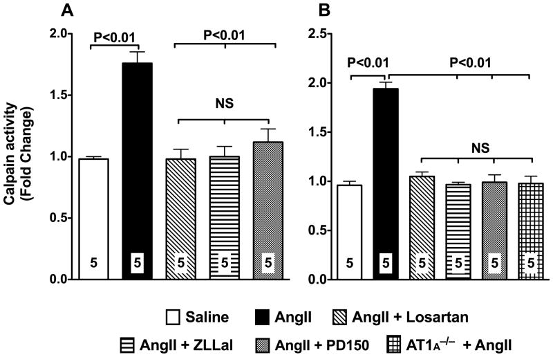
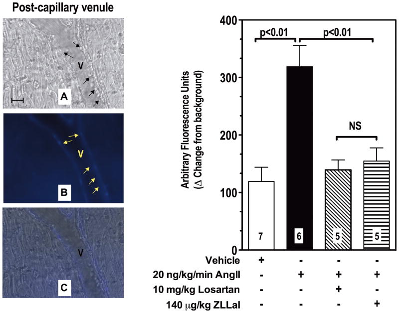
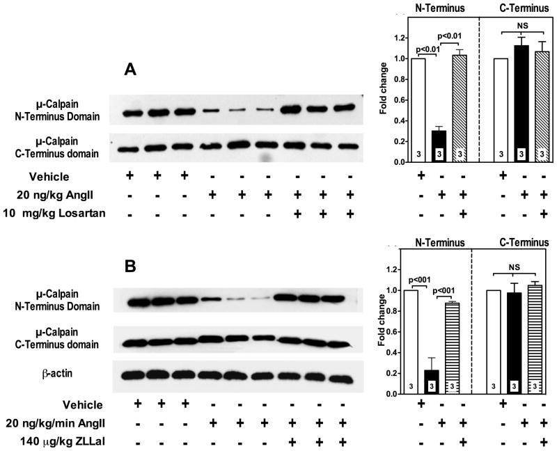
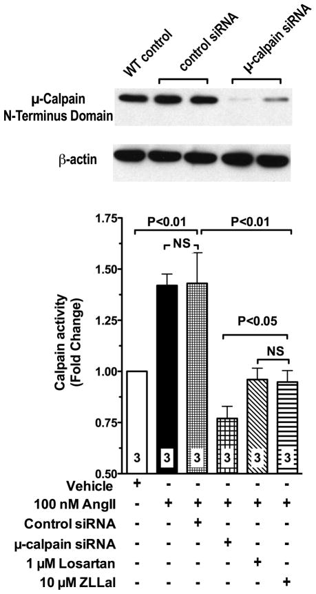
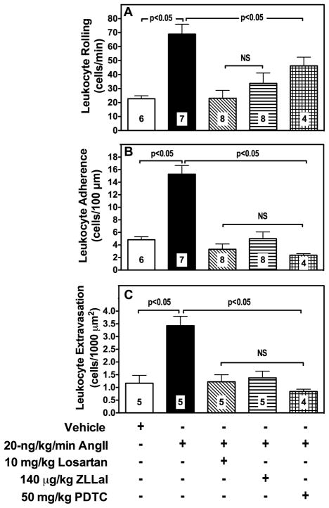
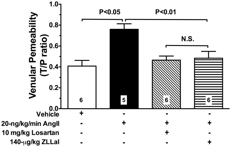
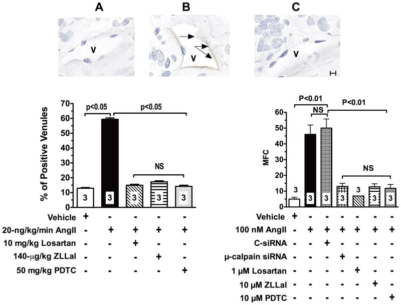
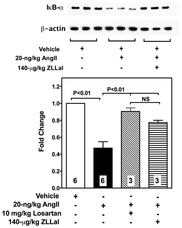
Methods and results: Subchronic administration of nonpressor doses of Ang II to rats and mice significantly increased vascular calpain activity via AT(1)R signaling. Intravital microscopy studies revealed that activation of vascular expressed calpains causes endothelial dysfunction with increased leukocyte-endothelium interactions and albumin permeability in the microcirculation. Western blot and immunohistochemistry studies confirmed that Ang II/AT(1)R signaling preferentially activates the constitutively expressed μ-calpain isoform and demonstrated a calpain-dependent degradation of IκBα, along with upregulation of nuclear factor κB-regulated endothelial cell adhesion molecules. These physiological and biochemical parameters were nearly normalized following inhibition of AT(1)R or calpain in vivo. RNA silencing studies in microvascular endothelial cells, along with knockout and transgenic mouse studies, further confirmed the role of μ-calpain in the endothelial adhesiveness induced by Ang II.

Conclusions: This study uncovers a novel role for calpain in the endothelial dysfunction of Ang II/AT(1)R signaling and establishes the calpain system as a novel molecular target of the vascular protective action of renin-angiotensin system inhibition. Our results may have significant clinical implications in vascular disease.


Figures

Figure 1
Figure 1. Activation of the AngII/AT1r signaling pathway increases vascular calpain activity
Calpain activity in vascular mesenteric extracts was measured using a fluorescent based assay with the use of the fluorescent probe T- Succ-LLVY-AMC. Two-week infusion of subpressor doses of AngII to rats (20 ng/Kg/min; left panel) or mice (500 ng/Kg/min; right panel) significantly increases calpain activity. A 5-day treatment of rat and mice with 10 mg/kg/day of the AT1r blocker losartan prevents upregulation of calpain activity in response to AngII. Similar results were obtained following treatment of AngII infused rats with two different calpain inhibitors, 140 μg/kg/day ZLLal or 1 mg/kg PD105. AngII failed to increase calpain activity in the vasculature of mice deficient in the AT1r. Data are expressed as the mean±SEM. Numbers at the base of the bars indicate the number of animals studied in each group.
Figure 2
Figure 2. Ang II Infusion increases leukocyte adhesion and calpain activity in post-capillary of the rat microcirculation
Post-capillary venules (V) were first viewed under brightfield microscopy to measure leukocyte rolling, adhesion and extravasation (panel A, black arrows). Subsequently they were superfused with the fluorogenic calpain substrate t-BOC-Leu-Met-CMAC to visualize and measure by computerized densitometry endothelial calpain activity in live rats (panel B, yellow arrows). Calpain activity was increased by AngII and nearly normalized by a five-day treatment with either the AT1r blocker losartan or the calpain inhibitor ZLLal. Noteworthy, increased leukocyte-endothelium interactions were observed in post-capillary venules experiencing increased calpain activity (overlay Panel C). Right graph shows quantitative analyses of calpain activity in all experimental groups of rat. Bars represent mean SEM, and numbers at the base of the bars represent the number of rats studied in each group.
Figure 3
Figure 3. AngII/AT1r signaling upregulates the μ-calpain isoform in mesenteric vascular extracts
μ-Calpain activity was assessed by immunoblot analysis using a primary antibody that recognizes the μ-calpain large subunit N-terminus domain, which is autolyzed in active calpains (panels A and B, upper lanes). Thus, calpain activation is demonstrated by loss of antibody recognition. A primary antibody against μ-calpain large subunit C-Terminus domain, which is stable and therefore recognizes both inactive and active μ-calpain, was used to quantify total μ-calpain content in the tissue extracts (panels A and B, middle lanes). Equal loading was also confirmed by β-actin staining (panel B, bottom lane). Bar graphs summarize densitometric analysis. AngII causes activation of μ-calpain as demonstrated by reduced N-Terminus domain expression (panels A and B, upper lanes). No significant changes were observed in the overall expression levels of μ-calpain in all groups of rats as demonstrated by lack of difference in C-Terminus domain expression (panels A and B, middle lanes). The effect of AngII on μ-calpain activation is reversed by a 5-day treatment with either the AT1r blocker losartan (panel A and upper graph) or the calpain inhibitor ZLLal (panel B and lower graph). Numbers at the base of the bars indicate the number of rats studied in each group.
Figure 4
Figure 4. Depletion of μ-calpain with siRNA prevents upregulation of calpain activity in MMEC endothelial cells exposed to AngII
Treatment of MMEC with siRNA to μ-calpain effectively reduces calpain expression levels (immunoblot). AngII fails to upregulates calpain activity in μ-calpain deficient MMEC (bar graph), thus demonstrating that the endothelial expressed μ-calpain isoform is the specific molecular target of AngII/AT1r signaling under our experimental conditions. AngII increases calpain activity via activation of the AT1r, as demonstrated by loss of increased calpain activity in MMEC treated with losartan. The calpain inhibitor ZLLal also prevented calpain activation in response to AngII. Calpain activity in attached MMEC was measured using a standard fluorescent assay and the calpain selective substrate T- Succ-LLVY-AMC. Numbers at the base of the bars indicate the number of independent experiments.
Figure 5
Figure 5. Leukocyte-endothelium interactions in mesenteric post-capillary venules
Leukocyte rolling (panel A), adhesion (panel B), and extravasation (panel C) were studied in all experimental groups of rats by brightfield intravital microscopy and expressed as the number of leukocytes/min, leukocytes/100-μm vessel length, and leukocytes/1000 μm2 extravascular space, respectively. AngII increases leukocyte endothelium interactions in the microcirculation, which were reduced similarly by losartan, ZLLal or PDTC treatment. Bars represent mean ± SEM, and numbers at the base of the bars represent the number of rats studied in each group.
Figure 6
Figure 6. Elevated AngII/AT1r signaling increases albumin permeability
The average permeability index to TR-labeled albumin was calculated during intravital microscopy in single mesenteric post-capillary venules of all experimental groups of rats. A two-week exposure of the mesenteric microcirculation to a low, subpressor dose of AngII increases albumin permeability via AT1r signaling and in a calpain-dependent manner. Thus, both losartan and the calpain inhibitor ZLLal attenuated albumin permeability in AngII injected rats. Data are expressed as the mean±SEM, and numbers at the base of the bars represent the number of rats studied in each group.
Figure 7
Figure 7. AngII upregulates expression levels of ICAM-1 in the vascular endothelium
In vivo. ICAM-1 expression in microvessels (V) of the mesentery was studied by immunohistochemistry in all experimental groups of rats. Representative photomicrographs (A, B and C) illustrate the effect of calpain inhibition on AngII-induced ICAM-1 expression in rat ileal venules (V). Brown immunoperoxidase reaction product (black arrows) indicates positive staining. Percentage of venules staining positive for ICAM-1 in all experimental groups of rats are illustrated in the bar graph to the left. Control microvessels had a very low level of ICAM-1 expression (Panel A and bar graph). ICAM-1 expression levels were markedly increased in AngII infused rats (Panel B, black arrows and bar graph). Losartan, ZLLal, and PDTC treatment returned ICAM-1 expression to control levels (Panel C and bar graph). In vitro. Right bar graph shows summary of ICAM-1 expression levels in mesenteric microvascular endothelial cells (MMEC) measured by flow cytometry. Upregulation of ICAM-1 in MMEC by AngII also occurs via activation of the AT1r and it is mediated by calpain activity as demonstrated by reduced ICAM-1 expression levels in MMEC treated with losartan, ZLLal, and PDTC respectively. Loss of AngII-induced upregulation of ICAM-1 following depletion of μ-calpain with siRNA confirms the role of the μ-calpain isoform in the process of ICAM-1 upregulation by AngII. MFC indicates mean fluorescence channels values for ICAM-1 staining measured by flow cytometry. Bars represent mean ± SEM, and numbers at the base of the bars represent either the number of rats studied in each group (left graph) or the number of independent experiments (right graph). Twenty tissue sections were studied in each rat to evaluate percentage of positive venules. Black line in Panel C represents a scale of 10 μm.
Figure 8
Figure 8. IκBα expression in the vasculature of AngII infused rats
IκBα levels in all experimental groups of rats were studied by western blot analysis in the cytosolic fractions of vascularized mesenteric tissue lysates (immunoblot). Bar graph shows densitometric analysis of IκBα expression levels in all experimental groups of rats. Reduced IκBα expression levels were found in the mesenteric vasculature of AngII-infused rats. Treatment of AngII-infused rats with either losartan or the calpain inhibitor ZLLal significantly restored IκB-α expression. Equal loading was confirmed by β-actin staining. Bars represent mean ± SEM, and numbers at the base of the bars represent the number of rats studied in each group.

References

    1. Fujitani K, Kambayashi J, Sakon M, Ohmi SI, Kawashima S, Yukawa M, Yano Y, Miyoshi H, Ikeda M, Shinoki N, Monden M. Identification of mu-, m-calpains and calpastatin and capture of mu-calpain activation in endothelial cells. Journal of cellular biochemistry. 1997;66:197–209. - PubMed
    1. Letavernier E, Perez J, Bellocq A, Mesnard L, de Castro Keller A, Haymann JP, Baud L. Targeting the calpain/calpastatin system as a new strategy to prevent cardiovascular remodeling in angiotensin ii-induced hypertension. Circulation research. 2008;102:720–728. - PubMed
    1. Jiang L, Wang M, Zhang J, Monticone RE, Telljohann R, Spinetti G, Pintus G, Lakatta EG. Increased aortic calpain-1 activity mediates age-associated angiotensin ii signaling of vascular smooth muscle cells. PLoS ONE. 2008;3:e2231. - PMC - PubMed
    1. Rateri DL, Moorleghen JJ, Balakrishnan A, Owens AP, 3rd, Howatt DA, Subramanian V, Poduri A, Charnigo R, Cassis LA, Daugherty A. Endothelial cell-specific deficiency of ang ii type 1a receptors attenuates ang ii-induced ascending aortic aneurysms in ldl receptor−/− mice. Circulation research. 2011 - PMC - PubMed
    1. Pastore L, Tessitore A, Martinotti S, Toniato E, Alesse E, Bravi MC, Ferri C, Desideri G, Gulino A, Santucci A. Angiotensin ii stimulates intercellular adhesion molecule-1 (icam-1) expression by human vascular endothelial cells and increases soluble icam-1 release in vivo. Circulation. 1999;100:1646–1652. - PubMed

Publication types

MeSH terms