. 2011 Jun;300(6):G1054-64.
doi: 10.1152/ajpgi.00055.2011. Epub 2011 Mar 17.

Occludin regulates macromolecule flux across the intestinal epithelial tight junction barrier

Affiliations

Occludin regulates macromolecule flux across the intestinal epithelial tight junction barrier

Rana Al-Sadi et al. Am J Physiol Gastrointest Liver Physiol. 2011 Jun.

Abstract

Defective intestinal epithelial tight junction (TJ) barrier has been shown to be an important pathogenic factor contributing to the development of intestinal inflammation. The expression of occludin is markedly decreased in intestinal permeability disorders, including in Crohn's disease, ulcerative colitis, and celiac disease, suggesting that the decrease in occludin expression may play a role in the increase in intestinal permeability. The purpose of this study was to delineate the involvement of occludin in intestinal epithelial TJ barrier by selective knock down of occludin in in vitro (filter-grown Caco-2 monolayers) and in vivo (recycling perfusion of mouse intestine) intestinal epithelial models. Our results indicated that occludin small-interfering RNA (siRNA) transfection causes an increase in transepithelial flux of various-sized probes, including urea, mannitol, inulin, and dextran, across the Caco-2 monolayers, without affecting the transepithelial resistance. The increase in relative flux rate was progressively greater for larger-sized probes, indicating that occludin depletion has the greatest effect on the flux of large macromolecules. siRNA-induced knock down of occludin in mouse intestine in vivo also caused an increase in intestinal permeability to dextran but did not affect intestinal tissue transepithelial resistance. In conclusion, these results show for the first time that occludin depletion in intestinal epithelial cells in vitro and in vivo leads to a selective or preferential increase in macromolecule flux, suggesting that occludin plays a crucial role in the maintenance of TJ barrier through the large-channel TJ pathway, the pathway responsible for the macromolecule flux.


Figures
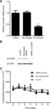
Fig. 1.
Fig. 1.
Effect of small-interfering RNA (siRNA)-induced knock down of occludin on filter-grown Caco-2 transepithelial electrical resistance (TER). A: siRNA occludin transfection caused a significant decrease in occludin mRNA levels as measured by real-time PCR (4 days posttransfection). *P < 0.001 vs. control. B: siRNA occludin transfection resulted in a near-complete depletion of occludin expression as assessed by Western blot analysis (6 days posttransfection). C: siRNA-induced knock down of occludin did not affect Caco-2 TER over the 6-day experimental period compared with controls.
Fig. 2.
Fig. 2.
Effect of siRNA-induced knock down of occludin on transepithelial flux of increasing-molecular-size paracellular markers across filter-grown Caco-2 monolayers. A: occludin depletion caused a 3- to 4-fold increase in urea flux (means ± SE, n = 6). *P < 0.001 vs. control. B and C: occludin depletion caused a 5- to 6-fold increase in mannitol and l-glucose flux (means ± SE, n = 6). *P < 0.001 vs. control. D and E: occludin depletion caused an ∼25-fold increase in inulin and 10 kDa (K) dextran (Dex) flux (means ± SE, n = 6). *P < 0.0005 vs. control. F: occludin depletion caused an ∼45-fold increase in 70 kDa dextran flux (means ± SE, n = 6). *P < 0.0001 vs. control. G: graph of molecular radius of paracellular markers vs. relative increase in flux rate following occludin siRNA transfection (relative correlation coefficient, r = 0.98).
Fig. 3.
Fig. 3.
Effect of occludin siRNA on various transmembrane tight junction (TJ) protein and mRNA expression. A: siRNA occludin caused an increase in claudin-2 protein expression but did not affect claudins-1, -3, -5, and -8 protein expression as assessed by Western blot analysis. B: occludin depletion by siRNA transfection in Caco-2 monolayers did not affect the mRNA levels of claudins-1, -3, -5, and -8 as assessed by real-time PCR (4 days posttransfection). C: claudin-2 mRNA levels were significantly increased in occludin siRNA-transfected Caco-2 monolayers (means ± SE, n = 4). *P < 0.001 vs. control.
Fig. 4.
Fig. 4.
The effect of occludin siRNA transfection on junctional localization of occludin and claudin-2 as determined by immunofluorescent antibody labeling and visualized by confocal microscope. A and B: immunostaining of occludin (green) and claudin-2 (red) in control Caco-2 monolayers. C: colocalization of occludin and claudin-2. D: siRNA-induced knock down of occludin in Caco-2 monolayer resulted in a significant depletion of the junctional localization of occludin. E: siRNA-induced knock down of occludin in Caco-2 monolayer caused an increase in claudin-2 intensity at the junctional localization. F: colocalization of occludin and claudin-2 following occludin siRNA transfection. Magnification, ×40.
Fig. 5.
Fig. 5.
Effect of siRNA-induced knock down of claudin-2 on transepithelial flux of varying-sized paracellular markers in occludin (Occ)-depleted Caco-2 monolayers. A and B: claudin-2 depletion did not affect the occludin siRNA-induced increase in flux rate of inulin and 70 kDa dextran (means ± SE, n = 4). *P < 0.0001 vs. control. C: claudin-2 depletion did not affect the occludin siRNA-induced increase in flux rate of mannitol (means ± SE, n = 6). *P < 0.0001 vs. control. D: claudin-2 depletion significantly prevented the occludin siRNA-induced increase in flux rate of urea (means ± SE, n = 6). *P < 0.0035 vs. control. **P < 0.0358 vs. control. #P < 0.0358 vs. occludin siRNA transfection.
Fig. 6.
Fig. 6.
Effect of selective siRNA-induced knock down of occludin in mouse intestine in vivo on mRNA and protein expression of TJ proteins in mouse intestinal tissue. A: occludin siRNA transfection resulted in a near-complete depletion of occludin expression in small intestinal tissue as assessed by Western blot analysis (3 days post-siRNA transfection). B: occludin siRNA transfection caused a significant decrease in occludin mRNA levels as measured by real-time PCR (means ± SE, n = 4). *P < 0.001 vs. control. C: occludin siRNA caused an increase in claudin-2 protein expression in mouse small intestinal tissue as assessed by Western blot analysis. D: claudin-2 mRNA levels were significantly increased in occludin siRNA transfected in mouse intestinal tissue (means ± SE, n = 4). *P < 0.001 vs. control. E: occludin siRNA did not affect claudins-1, -3, -5, and -8 protein expressions in mouse intestinal tissue as assessed by Western blot analysis. F: occludin depletion by siRNA transfection in mouse intestinal tissue did not affect the mRNA levels of claudins (Cld)-1, -3, -5, and -8 as assessed by real-time PCR (4 days posttransfection) (means ± SE, n = 5).
Fig. 7.
Fig. 7.
Effect of siRNA-induced knock down of occludin on enterocyte epithelial expression of occludin and claudin-2 in vivo. A pure population of villus enterocytes was isolated by laser capture microdissection (LCM). A: before and after LCM image of small intestinal mucosal surface. Arrows indicate the epithelial cells removed by LCM. B: occludin siRNA transfection caused a significant decrease in enterocyte occludin mRNA level as measured by real-time PCR (4 days posttransfection) (means ± SE, n = 4). *P < 0.001 vs. control. C: claudin-2 mRNA level was significantly increased in occludin siRNA-transfected enterocytes (means ± SE, n = 4). *P < 0.0001 vs. control.
Fig. 8.
Fig. 8.
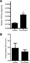
Effect of occludin siRNA in mouse small intestine permeability in vivo. A: occludin siRNA transfection in mouse intestine caused a significant increase in Texas red-labeled dextran (10 kDa) flux (means ± SE, n = 4). *P < 0.001 vs. control. B: occludin siRNA transfection did not affect the electrical resistance of small intestine tissue mounted on an Ussing chamber (means ± SE, n = 6).

References

    1. Al-Sadi R, Boivin M, Ma T. Mechanism of cytokine modulation of epithelial tight junction barrier. Front Biosci 14: 2765–2778, 2009 - PMC - PubMed
    1. Al-Sadi RM, Ma TY. IL-1beta causes an increase in intestinal epithelial tight junction permeability. J Immunol 178: 4641–4649, 2007 - PMC - PubMed
    1. Anderson JM, Van Itallie CM. Physiology and function of the tight junction. Cold Spring Harb Perspect Biol 1: a002584, 2009 - PMC - PubMed
    1. Arrieta MC, Madsen K, Doyle J, Meddings J. Reducing small intestinal permeability attenuates colitis in the IL10 gene-deficient mouse. Gut 58: 41–48, 2009 - PMC - PubMed
    1. Bamforth SD, Kniesel U, Wolburg H, Engelhardt B, Risau W. A dominant mutant of occludin disrupts tight junction structure and function. J Cell Sci 112: 1879–1888, 1999 - PubMed

Publication types

MeSH terms