. 2011 Oct;18(10):1573-83.
doi: 10.1038/cdd.2011.21. Epub 2011 Mar 18.

Phospholipase A(2) of peroxiredoxin 6 has a critical role in tumor necrosis factor-induced apoptosis

Affiliations

Phospholipase A(2) of peroxiredoxin 6 has a critical role in tumor necrosis factor-induced apoptosis

S Y Kim et al. Cell Death Differ. 2011 Oct.

Abstract

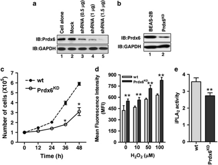
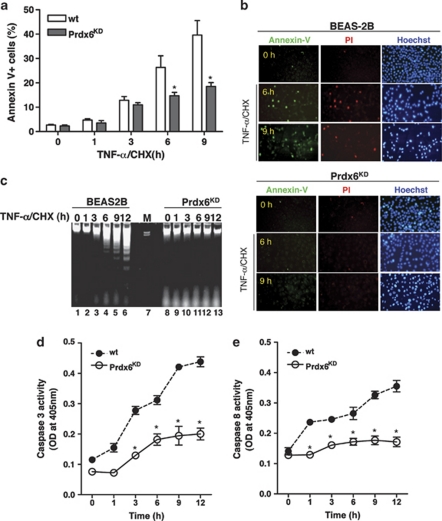
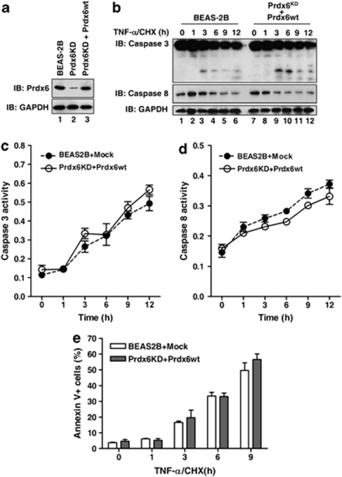
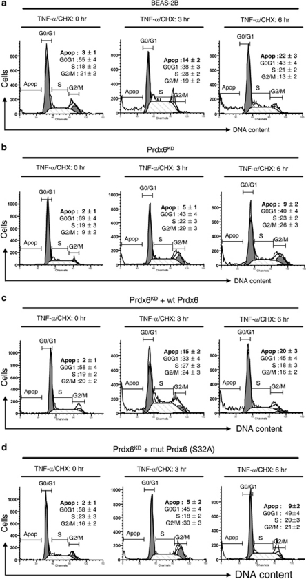
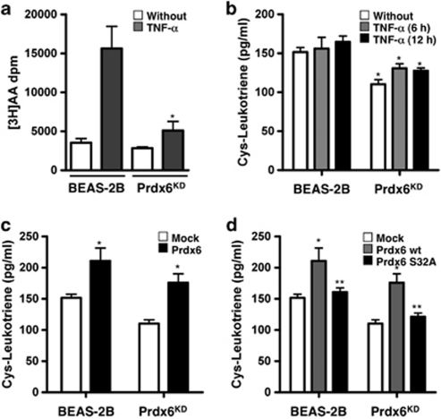
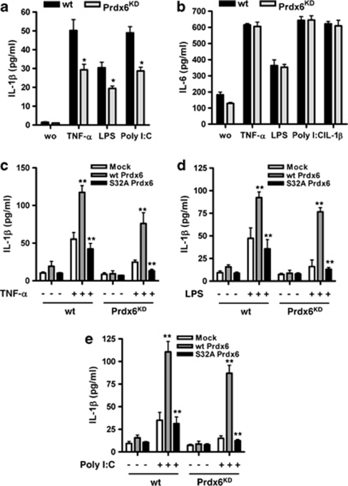
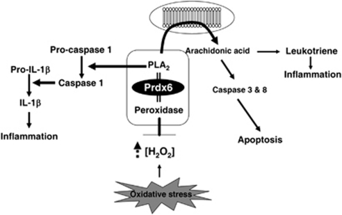
Peroxiredoxin 6 (Prdx6) is a bifunctional enzyme with peroxidase and phospholipase A(2) (PLA(2)) activities. Although the cellular function of the peroxidase of Prdx6 has been well elucidated, the function of the PLA(2) of Prdx6 is largely unknown. Here, we report a novel function for the PLA(2) in regulating TNF-induced apoptosis through arachidonic acid (AA) release and interleukin-1β (IL-1β) production. Prdx6 knockdown (Prdx6(KD)) in human bronchial epithelial cells (BEAS2B) shows severe decreases of peroxidase and PLA(2) activities. Surprisingly, Prdx6(KD) cells are markedly resistant to apoptosis induced by TNF-α in the presence of cycloheximide, but are highly sensitive to hydrogen peroxide-induced apoptosis. Furthermore, the release of AA and the production of IL-1β induced by proinflammatory stimuli, such as TNF-α, LPS, and poly I/C, are severely decreased in Prdx6(KD) cells. More interestingly, the restoration of Prdx6 expression with wild-type Prdx6, but not PLA(2)-mutant Prdx6 (S32A), in Prdx6(KD) cells dramatically induces the recovery of TNF-induced apoptosis, AA release, and IL-1β production, indicating specific roles for the PLA(2) activity of Prdx6. Our results provide new insights into the distinct roles of bifunctional Prdx6 with peroxidase and PLA(2) activities in oxidative stress-induced and TNF-induced apoptosis, respectively.


Figures

Figure 1
Figure 1
Prdx6KD BEAS-2B cells exhibit decreases of peroxidase and PLA2 activities. (a) Mock or pSuper.retro vector containing an shRNA specific for Prdx6 was transfected into BEAS-2B cells at different concentrations. The endogenous Prdx6 expression was evaluated. (b) Retroviruses containing an shRNA specific for Prdx6 were produced, as described in Materials and Methods. The cells were infected with the viruses and selected in puromycin-containing media for 30 days. The Prdx6-knockdown efficacy was examined. (c) In all, 1 × 10⁵ cells were plated into six wells and incubated for various times as indicated. At each different time, the cells were harvested and counted under trypan blue. (d) The wt BEAS-2B and Prdx6KD cells were exposed to different concentrations of H2O2, as indicated, for 2 h. The intracellular level of H2O2 was analyzed by DCF stain, as described in Materials and Methods. (e) The iPLA2 activities in wt BEAS-2B and Prdx6KD cells were measured according to the manufacturer's recommendations, as described in Materials and Methods. Bars represent mean±S.D. from at least three independent experiments. P-values were calculated using t-test versus wt BEAS-2B or Prdx6KD cells (*P<0.001 and **P<0.05)
Figure 2
Figure 2
Prdx6KD cells are highly susceptible to oxidative stress-induced apoptosis. (a) The wt BEAS-2B and Prdx6KD cells were exposed to different concentrations of H2O2 for 24 h. The cells were stained for AnnexinV. The percentage of AnnexinV-positive cells was analyzed with the FACSCalibur system and determined with the CellQuest software. The results are expressed as mean±S.D. for triplicate assays. (b) Prdx6KD cells were transfected with mock, wt Prdx6, and peroxidase-mutant Prdx6 (C47A) cells. The cells were exposed to 500 μ H2O2 for 24 h, and then AnnexinV+ cells were analyzed by flow cytometry. Bars represent mean±S.D. from at least three independent experiments. (c) The wt BEAS-2B and Prdx6KD cells were exposed to 500 μ H2O2 for 24 h. The cells were stained with AnnexinV/PI/Hoechst 33342, as described in Materials and Methods, and visualized with a fluorescence microscope. (d) The wt BEAS-2B and Prdx6KD cells were exposed to 500 μ H2O2 for various times as indicated. Cells were homogenized in 1 ml of lysis buffer. The genomic DNA extracts were prepared, as described in Materials and Methods, run on 1.8% agarose gels and visualized under UV illumination. (e and f) The wt BEAS-2B and Prdx6KD cells were exposed to 500 μ H2O2 for different times as indicated. Caspase-3 (e) and caspase-8 (f) activities were measured using the CaspACE kit according to the manufacturer's instructions. The results are expressed as mean±S.D. for triplicate assays. P-values were calculated using t-test versus wt BEAS-2B cells (*P<0.001 and **P<0.05)
Figure 3
Figure 3
Prdx6KD cells show strong resistance to TNF-α/CHX-induced apoptosis. (a) The wt BEAS-2B and Prdx6KD cells were treated with 100 ng/ml TNF-α/3 μ CHX for various times as indicated. The cells were stained for AnnexinV. The percentage of AnnexinV-positive cells was analyzed with the FACSCalibur system and determined with the CellQuest software. The results are expressed as mean±S.D. for triplicate assays. (b) The wt BEAS-2B and Prdx6KD cells were treated with TNF-α/CHX for 9 h. The cells were stained with AnnexinV/PI/Hoechst 33342, as described in Materials and Methods, and visualized with a fluorescence microscope. (c) The wt BEAS-2B and Prdx6KD cells were treated with TNF-α/CHX for various times as indicated. Cells were homogenized in 1 ml of lysis buffer. The genomic DNA extracts were prepared, as described in Materials and Methods, run on 1.8% agarose gels, and visualized under UV illumination. (d and e) The wt BEAS-2B and Prdx6KD cells were treated with TNF-α/CHX for different times as indicated. Caspase-3 (d) and caspase-8 (e) activities were measured using the CaspACE kit according to the manufacturer's instructions. The results are expressed as mean±S.D. for triplicate assays. P-values were calculated using t-test versus wt BEAS-2B (*P<0.05)
Figure 4
Figure 4
TNF-α/CHX-induced apoptosis was dramatic in Prdx6-overexpressing Prdx6KD cells. (a) Prdx6 was transfected into Prdx6KD cells. The expression of Prdx6 was compared in wt BEAS-2B, Prdx6KD, and Prdx6-overexpressing Prdx6KD cells with an antibody specific for Prdx6. (b) The wt BEAS-2B and Prdx6-overexpressing Prdx6KD cells were treated with TNF-α/CHX for various times as indicated. The lysates were examined by western blotting using anti-caspase-3, anti-caspase-8, and anti-GAPDH. (c and d) The wt BEAS-2B and Prdx6-overexpressed Prdx6KD cells were treated with TNF-α/CHX for different times as indicated. Caspase-3 (c) and caspase-8 (d) activities were measured using the CaspACE kit according to the manufacturer's instructions. The results are expressed as mean±S.D. for triplicate assays. (e) The wt BEAS-2B and Prdx6-overexpressed Prdx6KD cells were treated with TNF-α/CHX for various times as indicated. The cells were stained for AnnexinV. The percentage of AnnexinV-positive cells was analyzed with the FACSCalibur system and determined with the CellQuest software. The results are expressed as mean±S.D. for triplicate assays
Figure 5
Figure 5
The PLA2 activity of Prdx6 is essential for TNF-α/CHX-induced apoptosis. The wt Prdx6 and mutant Prdx6 (S32A) were expressed in Prdx6KD cells. The wt BEAS-2B (a), Prdx6 (b), wt Prdx6-overexpressing Prdx6KD (c), and mutant Prdx6 (S32A)-overexpressing Prdx6KD cells (d) were treated with TNF-α/CHX for different times as indicated. Cells were washed with HBSS, stained with PI, as described in Materials and Methods, and analyzed with the FACSCalibur system. The cell-cycle distributions were determined with the Modfit LT 3.0 software. The results are expressed as mean±S.D. for triplicate assays
Figure 6
Figure 6
The PLA2 activity of Prdx6 regulates the release of AA for the synthesis of leukotriene in response to TNF-α stimulation. (a) The wt BEAS-2B and Prdx6KD cells were incorporated with [3H] AA and stimulated with TNF-α, as described in Materials and Methods. The supernatants were collected from three separate wells. The samples were chromatographed by HPLC and the elutions were measured for radioactivity. The results are expressed as mean±S.D. for triplicate assays. (b) The wt BEAS-2B and Prdx6KD cells were stimulated with TNF-α for various times as indicated. The supernatants were collected, and then levels of cysteinyl leukotriene were measured with the Cysteinyl leukotriene Express EIA Kit (Cayman Chemical, Ann Arbor, MI, USA) according to the manufacturer's protocol. The results are expressed as mean±S.D. for triplicate assays (c) Mock or wt Prdx6 vector was transfected into wt BEAS-2B and Prdx6KD cells. After 48 h, supernatants were collected, and then levels of cysteinyl leukotriene were measured. The results are expressed as mean±S.D. for triplicate assays. (d) Mock, wt Prdx6, and Prdx6 S32A-mutant vectors were transfected into wt BEAS-2B and Prdx6KD cells. After 48 h, supernatants were collected, and then levels of cysteinyl leukotriene were measured. The results are expressed as mean±S.D. for triplicate assays. P-values were calculated using t-test versus wt BEAS-2B cells (*P<0.001 and **P<0.05)
Figure 7
Figure 7
The PLA2 activity of Prdx6 regulates the production of IL-1β in response to proinflammatory stimulation. (a and b) The wt BEAS-2B and Prdx6KD cells were stimulated with TNF-α, LPS, poly I/C, and IL-1β for 12 h. Supernatants were collected, and then levels of IL-1β (a) and IL-6 (b) were measured, as described in Materials and Methods. The results are expressed as mean±S.D. for triplicate assays. (c) Mock, wt Prdx6, and S32A Prdx6-mutant vectors were transfected into wt BEAS-2B and Prdx6KD cells. The cells were stimulated with TNF-α, and then the level of IL-1β was measured, as described in Materials and Methods. The results are expressed as mean±S.D. for triplicate assays. (d) Mock, wt Prdx6, and S32A Prdx6-mutant vectors were transfected into wt BEAS-2B and Prdx6KD cells. The cells were stimulated with LPS, and then the level of IL-1β was measured, as described in Materials and Methods. The results are expressed as mean±S.D. for triplicate assays. (e) Mock, wt Prdx6, and S32A Prdx6-mutant vectors were transfected into wt BEAS-2B and Prdx6KD cells. The cells were stimulated with poly I/C, and then the level of IL-1β was measured, as described in Materials and Methods. The results are expressed as mean±S.D. for triplicate assays. P-values were calculated using t-test versus wt BEAS-2B cells, mock-trasfected, or wt Prdx6-transfected cells (*P<0.05 and **P<0.001)
Figure 8
Figure 8
A possible model of cellular functions of bifunctional Prdx6 with peroxidase and PLA2 activities. The peroxidase of Prdx6 acts to eliminate the increased intracellular H2O2 induced by a variety of stimuli and thereby has a key role in the protection against cellular damages in oxidative stress, as shown in the current work and many previous reports. Other cellular functions of the PLA2 activity of Prdx6 may be involved in the TNF-induced apoptosis and the proinflammatory response. PLA2 induces the release of AA, which is involved in the inflammatory response by its metabolic mediator or in apoptosis linked with a caspase-dependent pathway. In addition, PLA2 induces the activation of IL-1β-converting enzyme and caspase-1, and in turn regulates the production of IL-β. IL-1β as a potential proinflammatory cytokine is involved in the inflammatory response

Similar articles

Cited by

References

    1. Rhee SG, Kang SW, Chang TS, Jeong W, Kim K. Peroxiredoxin, a novel family of peroxidases. IUBMB Life. 2001;52:35–41. - PubMed
    1. Woo HA, Chae HZ, Hwang SC, Yang KS, Kang SW, Kim K, et al. Reversing the inactivation of peroxiredoxins caused by cysteine sulfinic acid formation. Science. 2003;300:653–656. - PubMed
    1. Rhee SG, Chae HZ, Kim K. Peroxiredoxins: a historical overview and speculative preview of novel mechanisms and emerging concepts in cell signaling. Free Radic Biol Med. 2005;38:1543–1552. - PubMed
    1. Wood ZA, Poole LB, Karplus PA. Peroxiredoxin evolution and the regulation of hydrogen peroxide signaling. Science. 2003;300:650–653. - PubMed
    1. Chen JW, Dodia C, Feinstein SI, Jain MK, Fisher AB. 1-Cys peroxiredoxin, a bifunctional enzyme with glutathione peroxidase and phospholipase A2 Activities. J Biol Chem. 2000;275:28421–28427. - PubMed

Publication types

MeSH terms