

PSA-NCAM is Expressed in Immature, but not Recently Generated, Neurons in the Adult Cat Cerebral Cortex Layer II
- PMID: 21415912
- PMCID: PMC3042688
- DOI: 10.3389/fnins.2011.00017
PSA-NCAM is Expressed in Immature, but not Recently Generated, Neurons in the Adult Cat Cerebral Cortex Layer II
Abstract
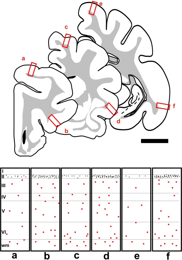
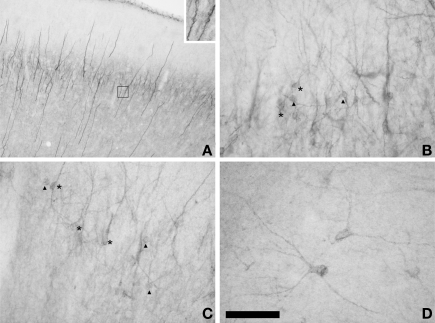
Neuronal production persists during adulthood in the dentate gyrus and the olfactory bulb, where substantial numbers of immature neurons can be found. These cells can also be found in the paleocortex layer II of adult rodents, but in this case most of them have been generated during embryogenesis. Recent reports have described the presence of similar cells, with a wider distribution, in the cerebral cortex of adult cats and primates and have suggested that they may develop into interneurons. The objective of this study is to verify this hypothesis and to explore the origin of these immature neurons in adult cats. We have analyzed their distribution using immunohistochemical analysis of the polysialylated form of the neural cell adhesion molecule (PSA-NCAM) and their phenotype using markers of mature neurons and different interneuronal populations. Additionally, we have explored the origin of these cells administering 5'bromodeoxyuridine (5'BrdU) during adulthood. Immature neurons were widely dispersed in the cerebral cortex layers II and upper III, being specially abundant in the piriform and entorhinal cortices, in the ventral portions of the frontal and temporoparietal lobes, but relatively scarce in dorsal regions, such as the primary visual areas. Only a small fraction of PSA-NCAM expressing cells in layer II expressed the mature neuronal marker NeuN and virtually none of them expressed calcium binding proteins or neuropeptides. By contrast, most, if not all of these cells expressed the transcription factor Tbr-1, specifically expressed by pallium-derived principal neurons, but not CAMKII, a marker of mature excitatory neurons. Absence of PSA-NCAM/5'BrdU colocalization suggests that, as in rats, these cells were not generated during adulthood. Together, these results indicate that immature neurons in the adult cat cerebral cortex layer II are not recently generated and that they may differentiate into principal neurons.
Keywords: adult neurogenesis; interneuron; neuronal differentiation; principal neuron; structural plasticity.
Figures


Similar articles
-
Adult neurogenesis and "immature" neurons in mammals: an evolutionary trade-off in plasticity?Brain Struct Funct. 2024 Nov;229(8):1775-1793. doi: 10.1007/s00429-023-02717-9. Epub 2023 Oct 13. Brain Struct Funct. 2024. PMID: 37833544 Free PMC article. Review.
-
Olfactory bulbectomy, but not odor conditioned aversion, induces the differentiation of immature neurons in the adult rat piriform cortex.Neuroscience. 2011 May 5;181:18-27. doi: 10.1016/j.neuroscience.2011.03.004. Epub 2011 Mar 5. Neuroscience. 2011. PMID: 21382447
-
Characterization and isolation of immature neurons of the adult mouse piriform cortex.Dev Neurobiol. 2016 Jul;76(7):748-63. doi: 10.1002/dneu.22357. Epub 2015 Oct 31. Dev Neurobiol. 2016. PMID: 26487449
-
Neurochemical Phenotype of Reelin Immunoreactive Cells in the Piriform Cortex Layer II.Front Cell Neurosci. 2016 Mar 10;10:65. doi: 10.3389/fncel.2016.00065. eCollection 2016. Front Cell Neurosci. 2016. PMID: 27013976 Free PMC article.
-
Distribution and possible roles of the highly polysialylated neural cell adhesion molecule (NCAM-H) in the developing and adult central nervous system.Neurosci Res. 1993 Sep;17(4):265-90. doi: 10.1016/0168-0102(93)90111-3. Neurosci Res. 1993. PMID: 8264989 Review.
Cited by
-
Chronic lead exposure reduces doublecortin-expressing immature neurons in young adult guinea pig cerebral cortex.BMC Neurosci. 2012 Jul 19;13:82. doi: 10.1186/1471-2202-13-82. BMC Neurosci. 2012. PMID: 22812564 Free PMC article.
-
Adult neurogenesis and "immature" neurons in mammals: an evolutionary trade-off in plasticity?Brain Struct Funct. 2024 Nov;229(8):1775-1793. doi: 10.1007/s00429-023-02717-9. Epub 2023 Oct 13. Brain Struct Funct. 2024. PMID: 37833544 Free PMC article. Review.
-
Cellular Plasticity in the Adult Murine Piriform Cortex: Continuous Maturation of Dormant Precursors Into Excitatory Neurons.Cereb Cortex. 2018 Jul 1;28(7):2610-2621. doi: 10.1093/cercor/bhy087. Cereb Cortex. 2018. PMID: 29688272 Free PMC article.
-
Sialic acids in the brain: gangliosides and polysialic acid in nervous system development, stability, disease, and regeneration.Physiol Rev. 2014 Apr;94(2):461-518. doi: 10.1152/physrev.00033.2013. Physiol Rev. 2014. PMID: 24692354 Free PMC article. Review.
-
Immature excitatory neurons develop during adolescence in the human amygdala.Nat Commun. 2019 Jun 21;10(1):2748. doi: 10.1038/s41467-019-10765-1. Nat Commun. 2019. PMID: 31227709 Free PMC article.
References
-
- Bonfanti L. (2006). PSA-NCAM in mammalian structural plasticity and neurogenesis. Prog. Neurobiol. 80, 129–164 - PubMed
-
- Cai Y., Xiong K., Chu Y., Luo D. W., Luo X. G., Yuan X. Y., Struble R. G., Clough R. W., Spencer D. D., Williamson A., Kordower J. H., Patrylo P. R., Yan X. X. (2009). Doublecortin expression in adult cat and primate cerebral cortex relates to immature neurons that develop into GABAergic subgroups. Exp. Neurol. 216, 342–356 - PMC - PubMed
-
- Friedman B., Price J. L. (1986). Age-dependent cell death in the olfactory cortex: lack of transneuronal degeneration in neonates. J. Comp. Neurol. 246, 20–31 - PubMed
LinkOut - more resources
Full Text Sources
Research Materials
Miscellaneous