. 2009 Oct 27;1(1):e2009006.
doi: 10.4084/MJHID.2009.006.

Iron metabolism in thalassemia and sickle cell disease

Affiliations

Iron metabolism in thalassemia and sickle cell disease

Raffaella Mariani et al. Mediterr J Hematol Infect Dis. .

Abstract

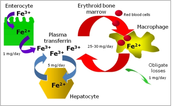
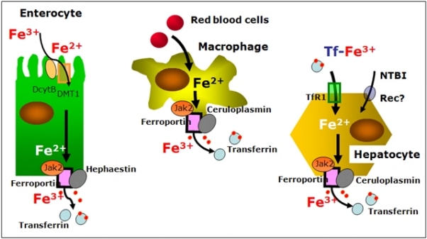
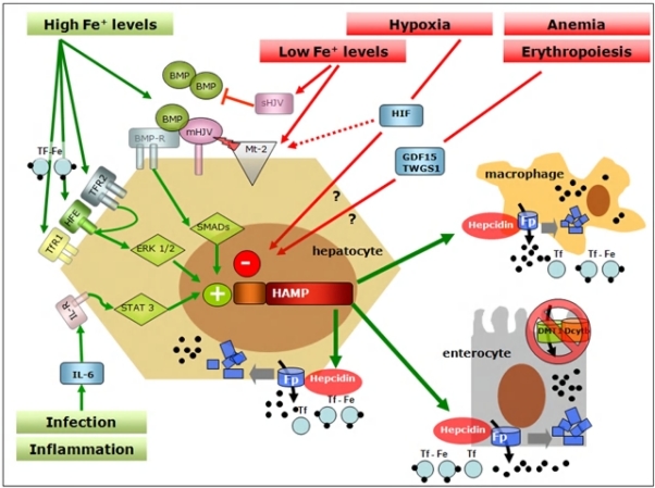
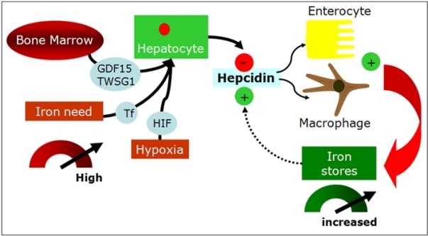
THERE ARE TWO MAIN MECHANISMS BY WHICH IRON OVERLOAD DEVELOPS IN THALASSEMIAS: increased iron absorption due to ineffective erythropoiesis and blood transfusions. In nontransfused patients with severe thalassemia, abnormal dietary iron absorption increases body iron burden between 2 and 5 g per year. If regular transfusions are required, this doubles the rate of iron accumulation leading to earlier massive iron overload and iron-related damage. Iron metabolism largely differs between thalassemias and sickle cell disease, but chronic transfusion therapy partially normalize many of the disparities between the diseases, making iron overload an important issue to be considered in the management of patients with sickle cell disease too. The present review summarizes the actual knowledge on the regulatory pathways of iron homeostasis. In particular, the data presented indicate the inextricably link between erythropoiesis and iron metabolism and the key role of hepcidin in coordinating iron procurement according to erythropoietic requirement. The role of erythropoietin, hypoxia, erythroid-dependent soluble factors and iron in regulating hepcidin transcription are discussed as well as differences and similarities in iron homeostasis between thalassemia syndromes and sickle cell disease.


Figures

Figure 1.
Figure 1.
Body iron homeostasis.
Figure 2.
Figure 2.
Cells regulating body-iron homeostasis. Enterocytes, macrophages and hepatocytes acquire iron from different sources and deliver it to the rest of the body through the iron exporter ferroportin, which needs copper-ferroxidases to release iron to plasma transferrin. Ferroportin acts under the control of hepcidin and this interaction can explain the systemic regulation of iron metabolism.
Figure 3.
Figure 3.
Signal pathways in systemic regulation of hepcidin. Many stimuli regulate expression of Hepcidin gene (HAMP) in the liver. One of the best known positive modulator is represented by Bone Morphogenetic Proteins (BMPs) that bind BMP-Receptor (BMP-R) on the surface of the hepatocyte resulting in SMAD-mediated induction of HAMP transcription. Hemojuvelin (mHJV) increases this signal acting as BMP co-receptor on the cell surface. In contrast, the soluble forms of HJV (sHJV), produced by HJV cleavage by furin at position 335, act as “decoy-receptor” competing with mHJV for the BMP ligand. Matriptase-2 (Mt2), which is activated by iron deficiency and by hypoxia, is the most potent inhibitor of hepcidin production by cleaving mHJV on hepatocyte surface and so preventing BMP-mediated hepcidin production. HAMP expression is also stimulated by inflammation, via the soluble mediator Interleukin-6 (IL6) and its specific membrane receptor (IL6-R) activating a STAT3-dependent signal pathway promoting HAMP transcription. HFE, TfR2 and TfR1 positively influence HAMP transcription in a ERK1/2 mediated way acting as a functional molecular complex on the cell surface playing a primary role in the hepatocyte sensing of circulating iron levels. Erythropoiesis, via the soluble mediator Growth Differentiation Factor 15 (GDF15) and Twisted Gastrulation (TWSG) 1, and hypoxia, via Hypoxia Inducible Factor (HIF), decrease HAMP expression (see text for further explanation).
Figure 4.
Figure 4.
Hepcidin regulation by erythroid- iron- and hypoxia-related signals in iron-loading anemias. (GDF= growth differentiation factor; TWSG= twisted gastrulation; TF= holo-transferrin; HIF= hypoxia inducible factor)

References

    1. Finch C. Regulators of iron balance in humans. Blood. 1994;84:1697–1702. - PubMed
    1. Fung EB, Harmatz P, Milet M, et al. Morbidity and mortality in chronically transfused subjects with thalassemia and sickle cell disease: A report from the multi-center study of iron overload. Am J Hematol. 2007;82:255–265. - PubMed
    1. De Domenico I, McVey Ward D, Kaplan J. Regulation of iron acquisition and storage: consequences for iron-linked disorders. Nat Rev Mol Cell Biol. 2008;9:72–81. - PubMed
    1. Andrews NC, Schmidt PJ. Iron homeostasis. Annu Rev Physiol. 2007;69:69–85. - PubMed
    1. Chasis JA, Mohandas N. Erythroblastic islands: niches for erythropoiesis. Blood. 2008;112:470–478. - PMC - PubMed

LinkOut - more resources