

Proteomic identification of biomarkers of vascular injury
- PMID: 21416056
- PMCID: PMC3056560
Proteomic identification of biomarkers of vascular injury
Abstract
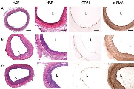
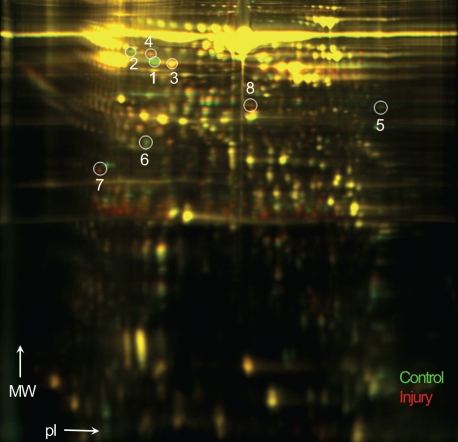
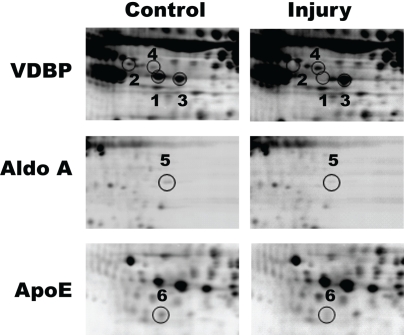
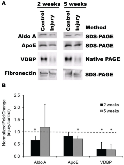
Predictive biomarkers may be beneficial for detecting, diagnosing, and assessing the risk of restenosis and vascular injury. We utilized proteomic profiling to identify protein markers in the blood following vascular injury, and corroborated the differential protein expression with immunological approaches. Rats underwent carotid artery injury, and plasma was collected after 2 or 5 weeks. Proteomic profiling was carried out by two-dimensional differential in-gel electrophoresis. The differentially expressed plasma proteins were identified by mass spectroscopy and confirmed by immunoblotting. Proteomic profiling by two-dimensional differential in-gel electrophoresis and mass spectroscopy revealed plasma proteins that were differentially expressed at 2 weeks after injury. Among the proteins identified included vitamin D binding protein (VDBP), aldolase A (aldo A), and apolipoproteinE (apoE). Immunoblotting results validated a significant reduction in these proteins in the plasma at 2 or 5 weeks after vascular injury, in comparison to control animals without vascular injury. These findings suggest that VDBP, aldo A, and apoE may be biomarkers for vascular injury, which will have important prognostic and diagnostic implications.
Keywords: Vascular injury; aldolase; angioplasty; apolipoprotein E; atherosclerosis; plasma marker; proteomic profiling; vitamin D binding protein.
Figures
Similar articles
-
Rationale and study design of the CardioGene Study: genomics of in-stent restenosis.Pharmacogenomics. 2004 Oct;5(7):952-1004. doi: 10.1517/14622416.5.7.949. Pharmacogenomics. 2004. PMID: 15469413
-
Two-Dimensional Differential Gel Electrophoresis to Identify Protein Biomarkers in Amniotic Fluid of Edwards Syndrome (Trisomy 18) Pregnancies.PLoS One. 2016 Jan 11;11(1):e0145908. doi: 10.1371/journal.pone.0145908. eCollection 2016. PLoS One. 2016. PMID: 26752631 Free PMC article.
-
Increased serum VDBP as a risk predictor for steroid resistance in asthma patients.Respir Med. 2016 May;114:111-6. doi: 10.1016/j.rmed.2016.03.011. Epub 2016 Mar 18. Respir Med. 2016. PMID: 27109820
-
Proteomic strategies in the search of new biomarkers in atherothrombosis.J Am Coll Cardiol. 2010 May 11;55(19):2009-16. doi: 10.1016/j.jacc.2010.01.036. J Am Coll Cardiol. 2010. PMID: 20447523 Review.
-
Proteomics for the identification of new prostate cancer biomarkers.Urol Oncol. 2006 May-Jun;24(3):231-6. doi: 10.1016/j.urolonc.2005.11.035. Urol Oncol. 2006. PMID: 16678055 Review.
Cited by
-
Differentiation of multipotent vascular stem cells contributes to vascular diseases.Nat Commun. 2012 Jun 6;3:875. doi: 10.1038/ncomms1867. Nat Commun. 2012. PMID: 22673902 Free PMC article.
-
cGMP inhibits TGF-beta signaling by sequestering Smad3 with cytosolic beta2-tubulin in pulmonary artery smooth muscle cells.Mol Endocrinol. 2011 Oct;25(10):1794-803. doi: 10.1210/me.2011-1009. Epub 2011 Aug 25. Mol Endocrinol. 2011. PMID: 21868450 Free PMC article.
-
Search for a diagnostic/prognostic biomarker for the brain cancer glioblastoma multiforme by 2D-DIGE-MS technique.Mol Cell Biochem. 2012 Aug;367(1-2):59-63. doi: 10.1007/s11010-012-1319-6. Epub 2012 May 1. Mol Cell Biochem. 2012. PMID: 22547198
-
Identification of the Transmembrane Glucose Regulated Protein 78 as a Biomarker for the Brain Cancer Glioblastoma Multiforme by Gene Expression and Proteomic Studies.J Membr Sci Technol. 2014 Feb 15;4(1):1000126. doi: 10.4172/2155-9589.1000126. J Membr Sci Technol. 2014. PMID: 26207187 Free PMC article.
-
Prevalence of ABCB4 polymorphisms in gallstone disease in han-Chinese population.Am J Transl Res. 2016 Feb 15;8(2):1218-27. eCollection 2016. Am J Transl Res. 2016. PMID: 27158408 Free PMC article.
References
-
- Rosamond W, Flegal K, Furie K, Go A, Greenlund K, Haase N, Hailpern SM, Ho M, Howard V, Kissela B, Kittner S, Lloyd-Jones D, McDermott M, Meigs J, Moy C, Nichol G, O'Donnell C, Roger V, Sorlie P, Steinberger J, Thom T, Wilson M, Hong Y. Heart disease and stroke statistics-2008 update: a report from the American Heart Association Statistics Committee and Stroke Statistics Subcommittee. Circulation. 2008;117:e25–146. - PubMed
-
- Glass CK, Witztum JL. Atherosclerosis. the road ahead. Cell. 2001;104:503–516. - PubMed
-
- Mateos-Caceres PJ, Garcia-Mendez A, Lopez Farre A, Macaya C, Nunez A, Gomez J, Alonso-Orgaz S, Carrasco C, Burgos ME, de Andres R, Granizo JJ, Farre J, Rico LA. Proteomic analysis of plasma from patients during an acute coronary syndrome. J Am Coll Cardiol. 2004;44:1578–1583. - PubMed
-
- Wilson AM, Kimura E, Harada RK, Nair N, Narasimhan B, Meng XY, Zhang F, Beck KR, Olin JW, Fung ET, Cooke JP. Beta2-microglobulin as a biomarker in peripheral arterial disease: proteomic profiling and clinical studies. Circulation. 2007;116:1396–1403. - PubMed
Grants and funding
LinkOut - more resources
Full Text Sources
Miscellaneous