

Topographic enhancement mapping of the cancer-associated breast stroma using breast MRI
- PMID: 21416100
- PMCID: PMC3698966
- DOI: 10.1039/c0ib00089b
Topographic enhancement mapping of the cancer-associated breast stroma using breast MRI
Abstract
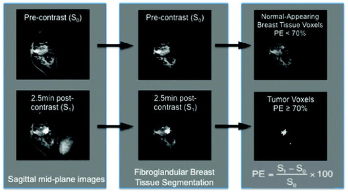
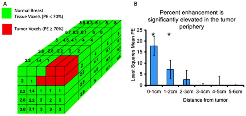
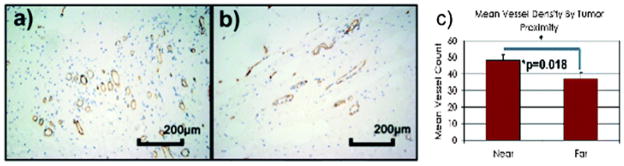
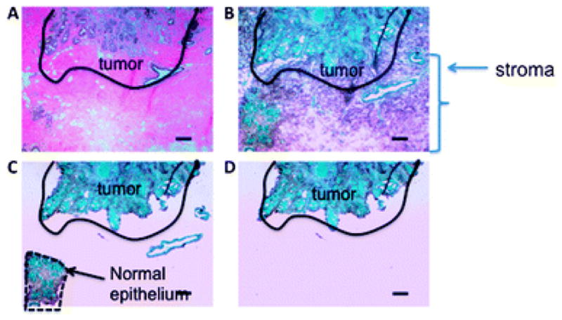
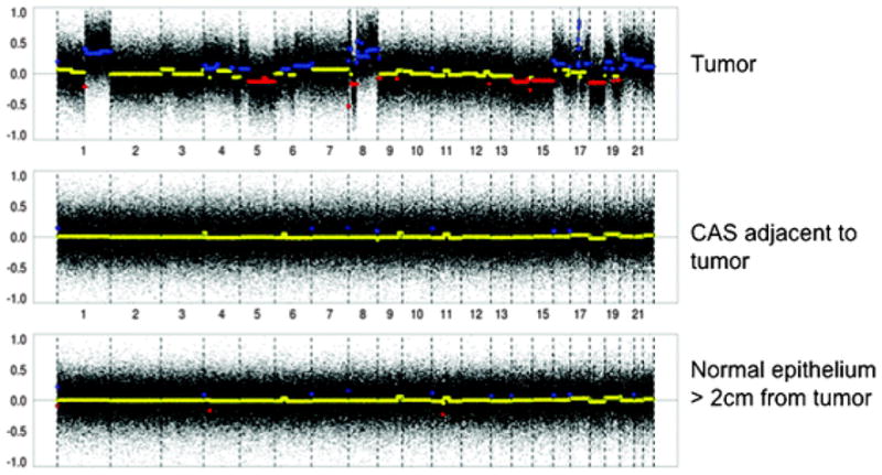
In animal and laboratory models, cancer-associated stroma, or elements of the supporting tissue surrounding a primary tumor, has been shown to be necessary for tumor evolution and progression. However, little is understood or studied regarding the properties of intact stroma in human cancer in vivo. In addition, for breast cancer patients, the optimal volume of local tissue to treat surrounding a primary tumor is not clear. Here, we performed an interdisciplinary study of normal-appearing breast tissue using breast magnetic resonance imaging (MRI), correlative histology and array comparative genomic hybridization to identify a cancer-associated stroma in humans. Using a novel technique for segmenting breast fibroglandular tissue, quantifiable topographic percent enhancement mapping of the stroma surrounding invasive breast cancer was found to be significantly elevated within 2 cm of the tumor edge. This region was also found to harbor increased microvessel density, and genomic changes that were closely associated with host normal breast tissue. These findings indicate that a cancer-associated stroma may be identified and characterized in human breast cancer using non-invasive imaging techniques. Identification of a cancer-associated stroma may be further developed to help guide local therapy to reduce recurrence and morbidity in breast cancer patients.
Figures
Similar articles
-
Correlation between mammographic density and volumetric fibroglandular tissue estimated on breast MR images.Med Phys. 2004 Apr;31(4):933-42. doi: 10.1118/1.1668512. Med Phys. 2004. PMID: 15125012 Clinical Trial.
-
Magnetic resonance imaging for secondary assessment of breast density in a high-risk cohort.Magn Reson Imaging. 2010 Jan;28(1):8-15. doi: 10.1016/j.mri.2009.05.040. Epub 2009 Jul 23. Magn Reson Imaging. 2010. PMID: 19631485 Free PMC article.
-
Breast stromal enhancement on MRI is associated with response to neoadjuvant chemotherapy.AJR Am J Roentgenol. 2008 Jun;190(6):1630-6. doi: 10.2214/AJR.07.2533. AJR Am J Roentgenol. 2008. PMID: 18492917 Clinical Trial.
-
Background parenchymal enhancement on breast MRI: A comprehensive review.J Magn Reson Imaging. 2020 Jan;51(1):43-61. doi: 10.1002/jmri.26762. Epub 2019 Apr 19. J Magn Reson Imaging. 2020. PMID: 31004391 Free PMC article. Review.
-
Dynamic contrast-enhanced breast MR imaging.Magn Reson Imaging Clin N Am. 2009 May;17(2):351-62. doi: 10.1016/j.mric.2009.01.010. Magn Reson Imaging Clin N Am. 2009. PMID: 19406363 Review.
Cited by
-
Features of MRI stromal enhancement with neoadjuvant chemotherapy: a subgroup analysis of the ACRIN 6657/I-SPY TRIAL.J Med Imaging (Bellingham). 2018 Jan;5(1):011014. doi: 10.1117/1.JMI.5.1.011014. Epub 2017 Dec 23. J Med Imaging (Bellingham). 2018. PMID: 29296631 Free PMC article.
-
MRI enhancement in stromal tissue surrounding breast tumors: association with recurrence free survival following neoadjuvant chemotherapy.PLoS One. 2013 May 7;8(5):e61969. doi: 10.1371/journal.pone.0061969. Print 2013. PLoS One. 2013. PMID: 23667451 Free PMC article.
-
Association of Peritumoral Radiomics With Tumor Biology and Pathologic Response to Preoperative Targeted Therapy for HER2 (ERBB2)-Positive Breast Cancer.JAMA Netw Open. 2019 Apr 5;2(4):e192561. doi: 10.1001/jamanetworkopen.2019.2561. JAMA Netw Open. 2019. PMID: 31002322 Free PMC article.
-
Prognostic Significance of CT-Attenuation of Tumor-Adjacent Breast Adipose Tissue in Breast Cancer Patients with Surgical Resection.Cancers (Basel). 2019 Aug 8;11(8):1135. doi: 10.3390/cancers11081135. Cancers (Basel). 2019. PMID: 31398863 Free PMC article.
-
Heterogeneous Enhancement Patterns of Tumor-adjacent Parenchyma at MR Imaging Are Associated with Dysregulated Signaling Pathways and Poor Survival in Breast Cancer.Radiology. 2017 Nov;285(2):401-413. doi: 10.1148/radiol.2017162823. Epub 2017 Jul 14. Radiology. 2017. PMID: 28708462 Free PMC article.
References
-
- Cunha GR, Hayward SW, Wang YZ. Role of stroma in carcinogenesis of the prostate. Differentiation. 2002;70:473–485. - PubMed
-
- Dolberg DS, Hollingsworth R, Hertle M, Bissell MJ. Wounding and its role in RSV-mediated tumor formation. Science. 1985;230:676–678. - PubMed
-
- Sieweke MH, Thompson NL, Sporn MB, Bissell MJ. Mediation of wound-related Rous sarcoma virus tumorigenesis by TGF-beta. Science. 1990;248:1656–1660. - PubMed
-
- Barcellos-Hoff MH, Ravani SA. Irradiated mammary gland stroma promotes the expression of tumorigenic potential by unirradiated epithelial cells. Cancer Res. 2000;60:1254–1260. - PubMed
-
- Deng G, Lu Y, Zlotnikov G, Thor AD, Smith HS. Loss of heterozygosity in normal tissue adjacent to breast carcinomas. Science. 1996;274:2057–2059. - PubMed
Publication types
MeSH terms
Grants and funding
LinkOut - more resources
Full Text Sources
Medical
