. 2011 Jul;39(7):1984-93.
doi: 10.1007/s10439-011-0293-6. Epub 2011 Mar 17.

Intertwined αβ spectrin meeting helical actin protofilament in the erythrocyte membrane skeleton: wrap-around vs. point-attachment

Affiliations

Intertwined αβ spectrin meeting helical actin protofilament in the erythrocyte membrane skeleton: wrap-around vs. point-attachment

Paul Sche et al. Ann Biomed Eng. 2011 Jul.

Abstract

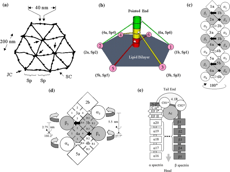
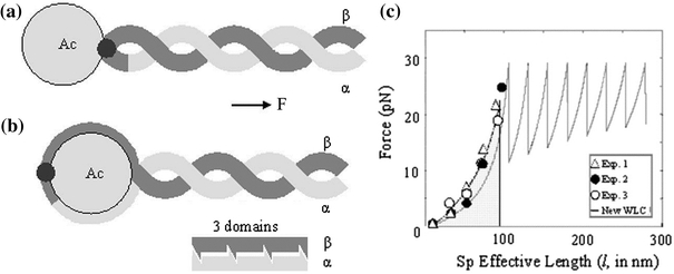
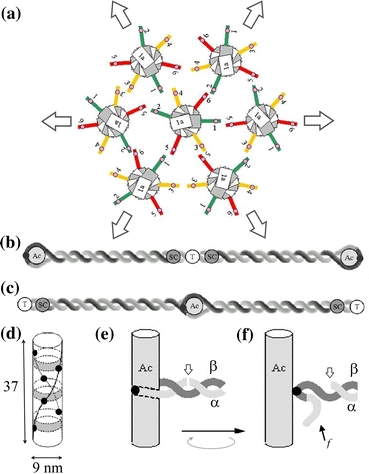
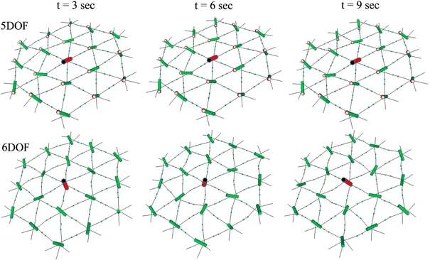
Our 3-D model for a junctional complex (JC) in the erythrocyte membrane skeleton proposed that the helical actin protofilament functions as a mechanical axis for three pairs of αβ spectrin (Sp), and each pair wraps around the protofilament in a back-to-back fashion. The distal end of each Sp is further associated with the lipid bilayer by a suspension complex (SC). Here, we detail how splitting and rejoining of αβ Sp around a protofilament may form a loop that sustains and equilibrates tension. Sequential association of β and α Sp solves the challenge of constructing multiple loops along the protofilament, and topological connection facilitates their re-association. The wrap-around model minimizes the strain of the actin binding site on β Sp due to tension, redirection, or sliding of intertwined Sp. Pairing Sp balances the opposing forces and provides a mechanism for elastic recovery. The wrap-around junction thus provides mechanical advantages over a point-attachment junction in maintaining the integrity and functionality of the network. Severing α or β Sp may convert a wrapping-around junction to a point-attachment junction. In that case, a "bow up" motion of JC during deformation may disturb or flip the overlaid lipid bilayer, and mark stressed erythrocytes for phagocytosis.


Figures

Figure 1
Figure 1
Sketches of the erythrocyte membrane skeleton and the wrap-around model from large to small scale. (a) The organization of the skeletal network, showing JC, SC, and Sp; (b) A 3-D view of a JC with a protofilament associated with three pairs of Sp and 6 SC, forming a basic repeating unit. (The angle between JC and the lipid bilayer is not specified); (c) A protofilament having 12 individual G actin and 6 occupied (filled) binding sites for β Sp; (d) An enlarged section of the protofilament bound with the middle pair: Sp3 (α3β3) and Sp4 (α4β4). Sp3 wraps around a pair of G actin (3a/3b); Sp4 wraps around the next pair of G actin (4a/4b) in the opposite direction (a–d modified from Ref. 27); (e) The detail sketch of α and β spectrin, which split and rejoin to wrap the protofilament. The tail end begins with the non-homologous domains of N-β, including ABD (CH1 and CH2), and that of C-α, including EFs. Cys (*) may form disulfide bonds. The dimer nucleation site spans α1-4 and β18-21. This sketch appeared in Ref. without detailed description. The size of linkers (# of residues): N-β (50), between CH1 and CH2 (10), between CH2 and β1 (19), between homologous β domains (8); C-α (75), between EF1 and EF2 (14), between EFII and α20 (8), and between α homologous domains (8). Molecules and domains are not to scale. The intertwined feature is not shown for clarity purposes
Figure 2
Figure 2
Point-attachment vs. wrap-around. (a) In a point-attachment junction, the ABD binds to a single point (solid circle) on the front side of the protofilament and force applied through Sp directly affects the binding. The unfolding (and open/close configurations) of domains would be the primary protective measure. (b) In a wrap-around junction, the ABD binds to the back side of the protofilament. α and β spectrin are arranged in an anti-parallel orientation and resists sliding through steric hindrance which is enhanced by intertwining. The greater the extension, the tighter the intercalating would be. (c) Modified force-extension curve of single stranded spectrin. The saw-tooth pattern represents successive unfolding of single spectrin repeats. The point-attachment junction features an irreversible drop (a vertical line) at the threshold at the point of detachment between Sp and protofilament. The lower panel shows 3 domains in each pitch
Figure 3
Figure 3
The conversion of a wrap-around junction to a point-attachment junction. (a) A two layer network (consisting of 1 CU and 6 PU) under equibiaxial extension. (b) A pair of SC at the center of a Sp tetramer. (c) A JC wrapped around by a pair of Sp (see Fig. 1d). (d) Side view of a JC, showing 6 occupied ABD (solid circles), 3 in each opposing helix, spiral from one end to the other. (e) One ABD in a wrap-around junction before a single strand cleavage (e.g., on α spectrin) (open arrow). (f) The cleavage may free the f segment from the protofilament and roll the protofilament during extension, converting a wrap-around junction to a point-attachment junction. AC: actin protofilament. T: Sp tetramerization site. f segment: a segment of single stranded spectrin between ABD and the cleavage site (may consist only β spectrin or a combination of α and β spectrin connected at the tail end)
Figure 4
Figure 4
Snap shots of a membrane skeletal network in 5DOF and 6DOF simulations during equibiaxial extension (λ = 1.3). Upper panels are 5DOF simulations (wrap-around junction) of a network with three layers at 3, 6, and 9 s; lower panels are their corresponding 6DOF simulations (point-attachment junction) based on Ref. . Note the “bow up” motion of the protofilament in CU (in red) in the 6DOF simulation. The point end of the protofilament is indicated by a black sphere (E-Tmod). See supplementary data for more information and complete animations

Similar articles

References

    1. Begg GE, Harper SL, Morris MB, Speicher DW. Initiation of spectrin dimerization involves complementary electrostatic interactions between paired triple-helical bundles. J. Biol. Chem. 2000;275(5):3279–3287. doi: 10.1074/jbc.275.5.3279. - DOI - PubMed
    1. Bratosin D, Mazurier J, Tissier JP, Slomianny C, Estaquier J, Russo-Marie F, Huart JJ, Freyssinet JM, Aminoff D, Ameisen JC, et al. Molecular mechanisms of erythrophagocytosis. Characterization of the senescent erythrocytes that are phagocytized by macrophages. C. R. Acad. Sci. III. 1997;320(10):811–818. - PubMed
    1. Byers TJ, Branton D. Visualization of the protein associations in the erythrocyte membrane skeleton. Proc. Natl Acad. Sci. USA. 1985;82(18):6153–6157. doi: 10.1073/pnas.82.18.6153. - DOI - PMC - PubMed
    1. Cherry L, Menhart N, Fung LW. Interactions of the alpha-spectrin N-terminal region with beta-spectrin Implications for the spectrin tetramerization reaction. J. Biol. Chem. 1999;274(4):2077–2084. doi: 10.1074/jbc.274.4.2077. - DOI - PubMed
    1. de Oliveira M, Vera C, Valdez P, Sharma Y, Skelton R, Sung LA. Nanomechanics of multiple units in the erythrocyte membrane skeletal network. Ann. Biomed. Eng. 2010;38(9):2956–2967. doi: 10.1007/s10439-010-0040-4. - DOI - PMC - PubMed

Publication types

LinkOut - more resources