

Crystal structure of tarocystatin-papain complex: implications for the inhibition property of group-2 phytocystatins
- PMID: 21416241
- PMCID: PMC3144364
- DOI: 10.1007/s00425-011-1398-8
Crystal structure of tarocystatin-papain complex: implications for the inhibition property of group-2 phytocystatins
Abstract
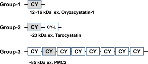
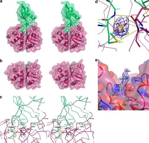
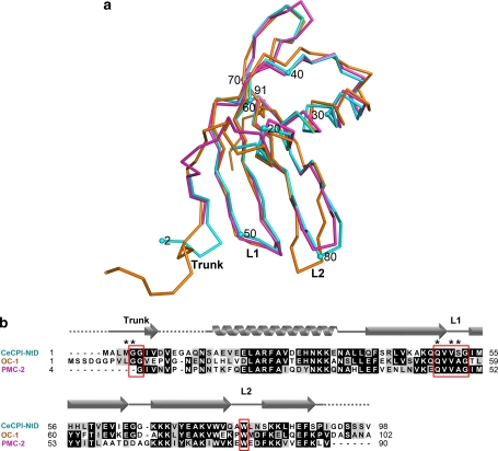
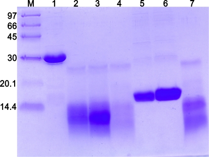
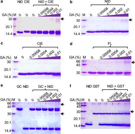
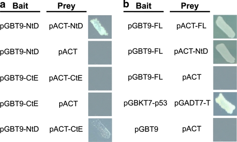
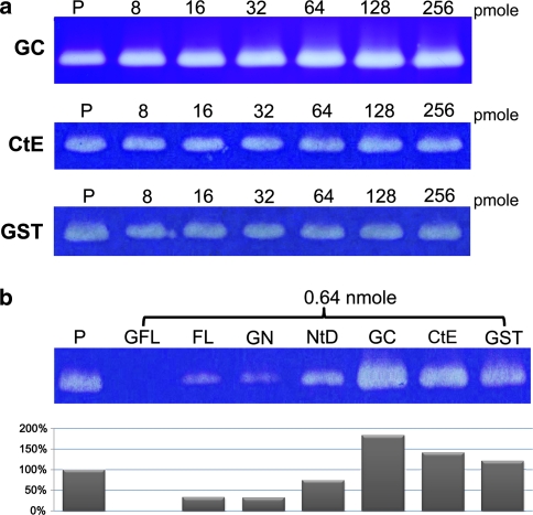
Tarocystatin (CeCPI) from taro (Colocasia esculenta cv. Kaohsiung no. 1), a group-2 phytocystatin, shares a conserved N-terminal cystatin domain (NtD) with other phytocystatins but contains a C-terminal cystatin-like extension (CtE). The structure of the tarocystatin-papain complex and the domain interaction between NtD and CtE in tarocystatin have not been determined. We resolved the crystal structure of the phytocystatin-papain complex at resolution 2.03 Å. Surprisingly, the structure of the NtD-papain complex in a stoichiometry of 1:1 could be built, with no CtE observed. Only two remnant residues of CtE could be built in the structure of the CtE-papain complex. Therefore, CtE is easily digested by papain. To further characterize the interaction between NtD and CtE, three segments of tarocystatin, including the full-length (FL), NtD and CtE, were used to analyze the domain-domain interaction and the inhibition ability. The results from glutaraldehyde cross-linking and yeast two-hybrid assay indicated the existence of an intrinsic flexibility in the region linking NtD and CtE for most tarocystatin molecules. In the inhibition activity assay, the glutathione-S-transferase (GST)-fused FL showed the highest inhibition ability without residual peptidase activity, and GST-NtD and FL showed almost the same inhibition ability, which was higher than with NtD alone. On the basis of the structures, the linker flexibility and inhibition activity of tarocystatins, we propose that the overhangs from the cystatin domain may enhance the inhibition ability of the cystatin domain against papain.
Figures
Similar articles
-
Characterization of inhibitory mechanism and antifungal activity between group-1 and group-2 phytocystatins from taro (Colocasia esculenta).FEBS J. 2008 Oct;275(20):4980-9. doi: 10.1111/j.1742-4658.2008.06631.x. Epub 2008 Sep 10. FEBS J. 2008. PMID: 18785929 Free PMC article.
-
Molecular cloning, recombinant gene expression, and antifungal activity of cystatin from taro (Colocasia esculenta cv. Kaosiung no. 1).Planta. 2005 Jun;221(4):493-501. doi: 10.1007/s00425-004-1462-8. Epub 2005 Jan 13. Planta. 2005. PMID: 15647900
-
Structural basis of the tarocystatin inhibitory mechanism against papain.Int J Biol Macromol. 2025 May;308(Pt 2):142647. doi: 10.1016/j.ijbiomac.2025.142647. Epub 2025 Mar 28. Int J Biol Macromol. 2025. PMID: 40158580
-
Structural and functional aspects of papain-like cysteine proteinases and their protein inhibitors.Biol Chem. 1997 Mar-Apr;378(3-4):141-50. Biol Chem. 1997. PMID: 9165064 Review.
-
Review: Unraveling the origin of the structural and functional diversity of plant cystatins.Plant Sci. 2022 Aug;321:111342. doi: 10.1016/j.plantsci.2022.111342. Epub 2022 May 27. Plant Sci. 2022. PMID: 35696902 Review.
Cited by
-
AlphaFold-Multimer predicts cross-kingdom interactions at the plant-pathogen interface.Nat Commun. 2023 Sep 27;14(1):6040. doi: 10.1038/s41467-023-41721-9. Nat Commun. 2023. PMID: 37758696 Free PMC article.
-
Phytocystatins: Defense Proteins against Phytophagous Insects and Acari.Int J Mol Sci. 2016 Oct 20;17(10):1747. doi: 10.3390/ijms17101747. Int J Mol Sci. 2016. PMID: 27775606 Free PMC article. Review.
-
A model of the C14-EPIC complex indicates hotspots for a protease-inhibitor arms race in the oomycete-potato interaction.Plant Signal Behav. 2011 Jan;6(1):109-12. doi: 10.4161/psb.6.1.14190. Epub 2011 Jan 1. Plant Signal Behav. 2011. PMID: 21301220 Free PMC article.
-
A novel cysteine proteinase inhibitor from seeds of Enterolobium contortisiliquum and its effect on Callosobruchus maculatus larvae.Biochem Biophys Rep. 2020 Dec 17;25:100876. doi: 10.1016/j.bbrep.2020.100876. eCollection 2021 Mar. Biochem Biophys Rep. 2020. PMID: 33364447 Free PMC article.
-
Arabidopsis thaliana Phytocystatin 6 Forms Functional Oligomer and Amyloid Fibril States.Biochemistry. 2023 Dec 5;62(23):3420-3429. doi: 10.1021/acs.biochem.3c00530. Epub 2023 Nov 21. Biochemistry. 2023. PMID: 37989209 Free PMC article.
References
-
- Barrett AJ. The cystatins: a diverse superfamily of cysteine peptidase inhibitors. Biomed Biochim Acta. 1986;45:1363–1374. - PubMed
Publication types
MeSH terms
Substances
Associated data
- Actions
- Actions
LinkOut - more resources
Full Text Sources
Research Materials
