Review
. 2011 Jul;462(1):119-34.
doi: 10.1007/s00424-011-0946-1. Epub 2011 Mar 18.

Cytoskeletal protein kinases: titin and its relations in mechanosensing

Affiliations
Review

Cytoskeletal protein kinases: titin and its relations in mechanosensing

Mathias Gautel. Pflugers Arch. 2011 Jul.

Abstract

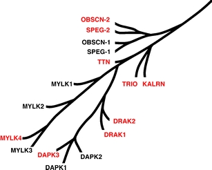
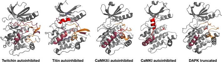
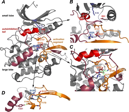
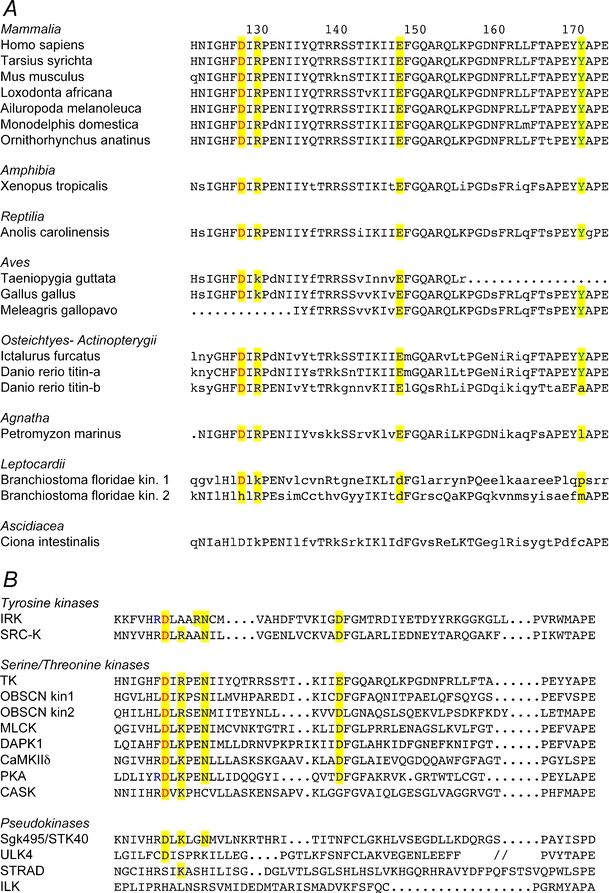
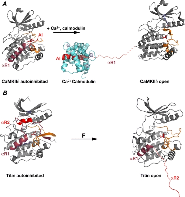
Titin, the giant elastic ruler protein of striated muscle sarcomeres, contains a catalytic kinase domain related to a family of intrasterically regulated protein kinases. The most extensively studied member of this branch of the human kinome is the Ca(2+)-calmodulin (CaM)-regulated myosin light-chain kinases (MLCK). However, not all kinases of the MLCK branch are functional MLCKs, and about half lack a CaM binding site in their C-terminal autoinhibitory tail (AI). A unifying feature is their association with the cytoskeleton, mostly via actin and myosin filaments. Titin kinase, similar to its invertebrate analogue twitchin kinase and likely other "MLCKs", is not Ca(2+)-calmodulin-activated. Recently, local protein unfolding of the C-terminal AI has emerged as a common mechanism in the activation of CaM kinases. Single-molecule data suggested that opening of the TK active site could also be achieved by mechanical unfolding of the AI. Mechanical modulation of catalytic activity might thus allow cytoskeletal signalling proteins to act as mechanosensors, creating feedback mechanisms between cytoskeletal tension and tension generation or cellular remodelling. Similar to other MLCK-like kinases like DRAK2 and DAPK1, TK is linked to protein turnover regulation via the autophagy/lysosomal system, suggesting the MLCK-like kinases have common functions beyond contraction regulation.


Figures

Fig. 1
Fig. 1
The MLCK branch of the human kinome (adapted from [70]). The CaM kinase family contains a group of related protein kinases that includes obscurin kinases 1 and 2 (OBSCN-1 and OBSCN-2), striated muscle preferentially expressed protein kinases 1 and 2 (SPEG-1 and SPEG-2), titin (TTN), trio and kalirin (KALRN), the myosin light-chain kinases of smooth muscle (MYLK1), skeletal muscle (MYLK2), cardiac muscle (MYLK3) and the more ubiquitously expressed MYLK4. Death-associated protein kinases 1–3 (DAPK1–3) and the death-associated protein kinase-related apoptosis-inducing protein kinase 1 and 2 (DRAK1 and DRAK2) form a separate sub-branch. Kinases that are unlikely to be CaM-regulated are shown in red
Fig. 2
Fig. 2
Structures of C-terminally autoinhibited protein kinases. Despite sequence identity of only about 40% and global overall structural similarity within the protein family, the titin and twitchin kinase domains show unique structural homology of their AI. In both kinases, the C-terminal autoinhibitory tail αR2 helix (bright red) tightly occludes the ATP binding cleft, while the αR1 helix (dark red in all kinases shown) does not directly block the catalytic cleft and shows a conserved topology. The function of the more C-terminal part of the autoinhibitory tail (bright red) is that of the actual autoinhibitor (in some cases like CaMKIIδ as a pseudosubstrate) despite very different secondary structure. In the case of CaMKIIδ and CaMKI, this segment contains the CaM binding site. Titin and twitchin share the unusual antiparallel β-sheet between the βR1 strand at the very C-terminus of the autoinhibitory tail and the activation segment βC10 strand. The complete autoinhibited structure of DAPK1 is not yet available; the published structure shows a similar αR1 helix topology as the other CaM kinase-like kinases; the CaM binding site is again in the—missing—C-terminal part of the autoinhibitory tail. Activation segments are shown in orange, the catalytic aspartate side chain in red. There are unresolved gaps in the CaMKI structure including a larger part of the activation segment. The following PDB entries are shown: nematode twitchin, 1KOA; human titin, 1TKI; human CaMKIIδ, 2VN9; human CaMKI, 2JC6; human DAPK1, A2A2
Fig. 3
Fig. 3
Structure of titin kinase and motifs involved in autoinhibition and catalysis. a Overall topology of TK, with the C-terminal autoinhibitory tail (hues of red) blocking access to the active site between the large and small lobes. The active site is blocked by αR2 (bright red). Side chains of key residues discussed are shown. The activation segment is shown in orange. b View towards the ATP binding cleft shows how αR2 (bright red) blocks ATP binding. K36, a key residue for coordinating the α/β-phosphates of ATP is shown as well as E51 in αC1. These two residues often form a salt bridge in active kinases but are separated by more than 4 Å in autoinhibited TK. c The catalysis loop around the catalytic base D127 shows the salt bridge and hydrogen bond network (green lines) between D127, R129, Q150 and the autoinhibitory Y170. d The tyrosine hydrogen bond network between the catalytic base D127, connecting Y170 and Y169 in the extended activation segment with αR1 in the autoinhibitory tail (dark red). Based on the crystal structure of TK, PDB entry 1TKI
Fig. 4
Fig. 4
a The catalysis loop and activation segment of titin kinase across the chordate classes. Key residues are highlighted in yellow, and residues identical to human titin are shown in capital letters. Note that the known avian titins show the lysine residue at position 129 that is canonical in other S/T kinases, where an arginine is conserved in all higher chordate titins down to lancelet. Note that sequence coverage makes the assessment of some amino acid exchanges in the lower species difficult. Available sequences suggest that the second kinase in the lancelet (Branchiostoma floridae) giant muscle protein (XP_002597967) is inactive, as the catalytic aspartate 127 (red) is exchanged for histidine. Numbering based on the TK crystal structure. b Catalysis loop and activation segments of exemplary human tyrosine and serine–threonine kinases and pseudokinases. Residues crucial for catalytic activity are highlighted in yellow, the catalytic aspartate is marked in red. Note the arginine residue at +2/4 from the catalytic aspartate instead of lysine in tyrosine kinases, a constellation found in TK and the obscurin kinase-2 domain. The boxed asparagine and acidic residues at D + 5 and in the DFG motif at the beginning of the activation segment are involved in coordinating the Mg2+ of Mg-ATP [120]. CASK, also sometimes classified as a pseudokinase [14], shows Mg2+-independent activity due to several mutations in the Mg2+-coordinating residues [81]. On this basis, the presumed pseudokinase STK40 might also show unusual catalytic activity like other pseudokinases [121]. ULK4, STRAD4 [102] and ILK [133], however, all lack crucial residues for catalysis, notably the catalytic aspartate for STRAD and ILK, and are inactive. Acronyms not mentioned in Fig. 1. IRK, insulin receptor kinase; SRC-K, Src kinase; PKA, cAMP-dependent protein kinase; CASK CaM-activated serine–threonine kinase [81]; STK40 serine–threonine kinase 40; ULK4, unc-51-like kinase 4; STRAD, STE20-related kinase adapter protein beta; ILK, integrin receptor-linked kinase
Fig. 5
Fig. 5
Local protein unfolding and refolding in the activation of protein kinases. a The genuinely CaM-activated CaMKIIδ is autoinhibited by the C-terminal autoinhibitory tail (hues of red) that blocks predominantly the catalytic base via an unstructured segment bearing the autoinhibitory threonine 287 (side chain shown in red). The activation segment (orange) is in an open configuration, similar to titin. Upon binding of CaM, the C-terminal autoinhibitory tail (hues of red) undergoes major unfolding–refolding events: the main inhibitory region around T287 (AI, bright red) adopts a helical conformation in complex with CaM, whereas the previously helical region around αR1 (dark red) unfolds and opens up further regulatory threonine phosphorylation sites [103]. b In titin kinase, external force (F) can lead to the activation-related unfolding of the autoinhibitory tail, with the main autoinhibitory αR2 (bright red) being pulled out of the ATP binding site and thus exposing D127 (red) and Y170 (orange). The αR1 helix (dark red) remains structured but eventually also unfolds; whether it undergoes conformational changes upon nbr1 binding is currently unknown. Open structures based on the low-velocity force-probe molecular dynamics simulations in [38], and the CaMKIId -CaM complex [103], PDB 2WEL. The unresolved segment in the CaMKIIδ AI structure has been extrapolated as a dashed red line. Autoinhibited structures as in Fig. 2.
Fig. 6
Fig. 6
Known interactions of the titin kinase associated, ubiquitin- and LC3-binding proteins Nbr1 and SQSTM1/p62. Both Nbr1 and SQSTM1 bind polyubiquitin chains via their C-terminal UBA domain and to the autophagy membrane component Atg8/LC3. Further links to small protein modifiers exist via the MURFs to SUMO [21, 79, 95]. Nbr1 and SQSTM1 interact with several other protein kinases including the atypical protein kinase-Cζp38 MAP kinase and several signalling adaptors, many of which are relevant for the control of muscle growth and remodelling
Fig. 7
Fig. 7
Sequence comparison of the autoinhibitory tails of the MLCK-like kinase branch. The CaM binding site in the skeletal MLCK M13 peptide [53, 89] and in confirmed and predicted CaM-regulated kinases are boxed in red; CaM kinase-I (CaMKI) is shown for comparison. About half of the MLCK-like kinases do not show a basic amphipathic region or a sufficiently long autoinhibitory tail and are thus possibly regulated by different mechanisms than CaM binding. The αR1 helix in titin and nematode twitchin are marked in magenta; αR2 (which blocks the ATP binding site) and βR1 are shown in red and blue, respectively. The synthetic titin peptide adopting helical conformation upon CaM binding [4], and the S100 binding peptide in twitchin [45] are underlined. Abbreviations as in Fig. 1.
Fig. 8
Fig. 8
Domain pattern and interactions of the MLCK-like kinases. This kinase family is highly modular, combining catalytic kinase domains with various cytoskeleton-associated domains of the intracellular immunoglobulin and fibronectin family, as well as spectrin and ankyrin repeat domains. Most, if not all, MLCK-like kinases are cytoskeleton-associated via interactions with components of the actin or myosin filaments (red). The exact topology of the actin and myosin binding sites in invertebrate twitchin has not yet been established (red italics). Titin as well as three genuine MLCKs contain an entropic spring sequence, the PEVK sequence (after the predominant amino acids: proline, glutamate, valine and lysine [63]), which forms an elastic connection between the N-terminal actin and C-terminal myosin associated regions. Obscurin, kalirin and trio are unique in that they combine cytoskeleton-associated Rho-GDP/GTP exchange factor domains with protein kinase domains. The presence of two tandem kinase domains in SPEG and obscurin seems specific for striated muscle proteins, but their function is unknown. The domain patterns of the giant titin, twitchin and obscurin proteins is shown only partially around the signalling domains and schematically around the PEVK segment for titin

References

    1. Agarkova I, Perriard JC. The M-band: an elastic web that crosslinks thick filaments in the center of the sarcomere. Trends Cell Biol. 2005;15:477–85. - PubMed
    1. Agarkova I, Ehler E, Lange S, Schoenauer R, Perriard JC. M-band: a safeguard for sarcomere stability? J Muscle Res Cell Motil. 2003;24:191–203. - PubMed
    1. Akiyama N, Ohnuki Y, Kunioka Y, Saeki Y, Yamada T. Transverse stiffness of myofibrils of skeletal and cardiac muscles studied by atomic force microscopy. J Physiol Sci. 2006;56:145–151. - PubMed
    1. Amodeo P, Castiglione Morelli MA, Strazzullo G, Fucile P, Gautel M, Motta A. Kinase recognition by calmodulin: modeling the interaction with the autoinhibitory region of human cardiac titin kinase. J Mol Biol. 2001;306:81–95. - PubMed
    1. Avrova SV, Shelud'ko NS, Borovikov YS, Galler S. Twitchin of mollusc smooth muscles can induce “catch”-like properties in human skeletal muscle: support for the assumption that the “catch” state involves twitchin linkages between myofilaments. J Comp Physiol B. 2009;179:945–50. - PubMed

Publication types

MeSH terms

LinkOut - more resources