Case Reports
. 2011 Apr;69(4):712-20.
doi: 10.1002/ana.22264. Epub 2011 Mar 17.

Familial prion disease with Alzheimer disease-like tau pathology and clinical phenotype

Affiliations
Case Reports

Familial prion disease with Alzheimer disease-like tau pathology and clinical phenotype

Suman Jayadev et al. Ann Neurol. 2011 Apr.

Abstract

Objective: To describe the Alzheimer disease (AD)-like clinical and pathological features, including marked neurofibrillary tangle (NFT) pathology, of a familial prion disease due to a rare nonsense mutation of the prion gene (PRNP).

Methods: Longitudinal clinical assessments were available for the proband and her mother. After death, both underwent neuropathological evaluation. PRNP was sequenced after failure to find immunopositive Aβ deposits in the proband and the documentation of prion protein (PrP) immunopositive pathology.

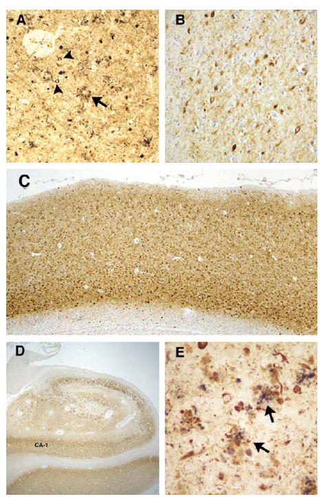
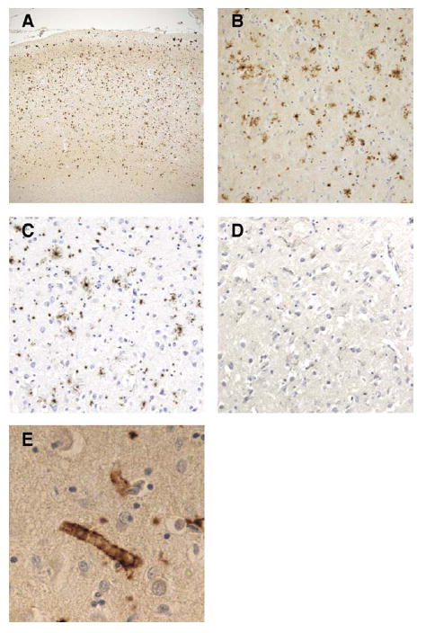
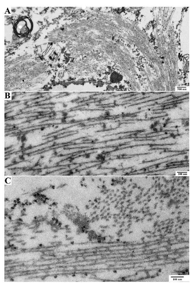
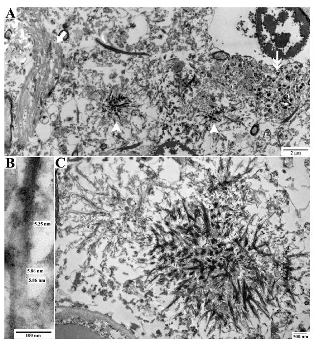
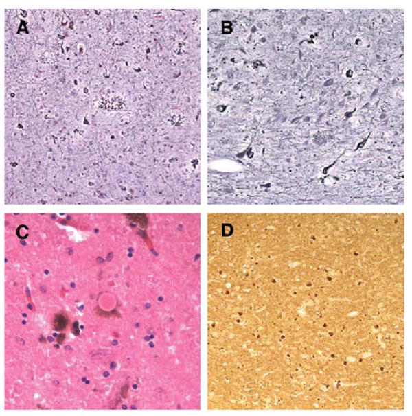
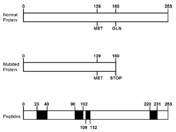
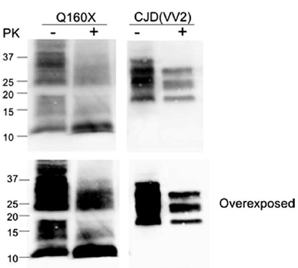
Results: The proband presented at age 42 years with a 3-year history of progressive short-term memory impairment and depression. Neuropsychological testing found impaired memory performance, with relatively preserved attention and construction. She was diagnosed with AD and died at age 47 years. Neuropathologic evaluation revealed extensive limbic and neocortical NFT formation and neuritic plaques consistent with a Braak stage of VI. The NFTs were immunopositive, with multiple tau antibodies, and electron microscopy revealed paired helical filaments. However, the neuritic plaques were immunonegative for Aβ, whereas immunostaining for PrP was positive. The mother of the proband had a similar presentation, including depression, and had been diagnosed clinically and pathologically as AD. Reevaluation of her brain tissue confirmed similar tau and PrP immunostaining findings. Genetic analysis revealed that both the proband and her mother had a rare PRNP mutation (Q160X) that resulted in the production of truncated PrP.

Interpretation: We suggest that PRNP mutations that result in a truncation of PrP lead to a prolonged clinical course consistent with a clinical diagnosis of AD and severe AD-like NFTs.


Conflict of interest statement

Potential Conflicts of Interest T.D.B. has received speaking fees and royalties from Athena Diagnostics.

Figures

FIGURE 1
FIGURE 1
Silver stain and tau pathology in proband (A). Bielschowsky silver stained sections from the proband demonstrate significant neurofibrillary tangle (arrowheads) and neuritic plaque (arrow) formation in the frontal cortex. Note the coreless plaque (arrow). Tau immunohistochemistry revealed severe immunopositive staining of neurofibrillary pathology in the frontal cortex (B and C, PHF1 antibody) and hippocampus (D, PHF-1 antibody). (E) Double immunolabeling of plaquelike structures in the hippocampus with prion protein (3F4, purple) and tau (PHF-1, brown) antibodies revealed a close anatomic relationship between these pathologic changes (arrows). Original magnification, A and B, ×63; C, ×13; D, ×7; E, ×126.
FIGURE 2
FIGURE 2
Prion protein (PrP) immunohistochemistry in frontal cortex of the proband. (A, B) PrP immunohistochemistry demonstrates widespread PrP deposition in frontal cortex. (C) Antibody specific to the amino acids 90 to 102 of PrP demonstrates immunopositive PrP deposits; (D) however, this was not observed with an antibody to amino acids 220 to 231 of the PrP. (E) PrP immunopositive angiopathy was also observed. Original magnification, A, ×13; B, C, and D, ×32; E, ×126.
FIGURE 3
FIGURE 3
Electron microscopy of neurofibrillary tangles in proband. Electron microscopic pictures of neuronal cytoplasm show the presence of neurofibrillary tangles. (A) Cytoplasm of a nerve cell containing a neurofibrillary tangle. (B, C) High-power image of neurofibrillary tangle shows paired helical filaments seen longitudinally (lower portion of the image) and in cross-section (upper right portion of image).
FIGURE 4
FIGURE 4
Electron microscopy of prion protein (PrP) deposition in proband. Electron microscopic pictures of neuropathologic lesions in the neuropil. (A) The neuropil shows a neurofibrillary tangle on the left (arrow), PrP amyloid deposits in the center (arrowheads), and a dystrophic neurite on the right (double arrow). (B) High-power image of filaments from a PrP amyloid plaque. (C) Starlike PrP amyloid plaques.
FIGURE 5
FIGURE 5
Histologic pathology in the proband’s mother. Holmes silver staining revealed severe neurofibrillary tangles and neuritic plaque pathology in the mother’s frontal cortex (A) and hippocampus (B). In addition, both classic Lewy bodies (C) and alpha-synuclein immunopositive inclusions and neurites (D) were observed. Original magnification, A and B, ×63; C, ×126; D, ×32.
FIGURE 6
FIGURE 6
Schematic representation of normal prion protein and truncated prion protein in this family. The scheme of the prion protein highlights the regions that recognized the antibodies used in the present study.
FIGURE 7
FIGURE 7
Western blot from proband brain tissue. Western blot analysis of enriched fractions of prion protein scrapie isoform (PrPSc) from brain samples of the PRNP-Q160X patient and a sporadic Creutzfeldt-Jakob disease (sCJD) subject with the 129VV genotype and type 2 proteinase K (PK)-resistant PrPSc (unglycosylated fragment ~19kDa) is shown. This enrichment reveals 2 major fractions of PrP that correspond to residual nonmutated (25–35kDa) PrP and full-length and cleaved (smear between 11 and 18kDa) mutated PrP. The predicted size of mature PrP-Q160X is ~17–18kDa, represented by the top of the second major fraction, with smaller fragments likely representing endogenously cleaved products of the PrP-Q160X mutated protein, the most prominent of which is ~11kDa. None of these smaller fractions is present in sCJD, even with overexposure, suggesting that they result directly from mutated PrP. Following PK treatment (+) the ~11kDa fragment remains most prominent. The smear of PK-resistant PrP that ranges from ~21 to 35kDa may represent PrP-Q160X oligomers or nonmutated PrP converted to PK-resistant fragments by PrP-Q160X. Blots were probed with antihuman PrP monoclonal antibody 3F4.

References

    1. Ironside JW, Ghetti B, Head MW. Prions diseases. In: Love S, Louis DN, Ellison DW, editors. Greenfield’s neuropathology. London, UK: Hodder Arnold; 2008. pp. 1197–1274.
    1. Kong Q, Suresicz WK, Petersen RB, et al. Inherited prion diseases. In: Prusiner SB, editor. Prion biology and diseases. 2. Cold Spring Harbor, NY: Cold Spring Harbor Laboratory Press; 2004. pp. 673–775.
    1. Finckh U, Muller-Thomsen T, Mann U, et al. High prevalence of pathogenic mutations in patients with early-onset dementia detected by sequence analyses of four different genes. Am J Hum Genet. 2000;66:110–117. - PMC - PubMed
    1. Ghetti B, Piccardo P, Spillantini MG, et al. Vascular variant of prion protein cerebral amyloidosis with tau-positive neurofibrillary tangles: the phenotype of the stop codon 145 mutation in PRNP. Proc Natl Acad Sci U S A. 1996;93:744–748. - PMC - PubMed
    1. Piccardo P, Ghetti B, Dickson DW, et al. Gerstmann-Straussler-Scheinker disease (PRNP P102L): amyloid deposits are best recognized by antibodies directed to epitopes in PrP region 90-165. J Neuropathol Exp Neurol. 1995;54:790–801. - PubMed

Publication types

MeSH terms