

Regional brain volume reductions relate to facial dysmorphology and neurocognitive function in fetal alcohol spectrum disorders
- PMID: 21416562
- PMCID: PMC3812802
- DOI: 10.1002/hbm.21260
Regional brain volume reductions relate to facial dysmorphology and neurocognitive function in fetal alcohol spectrum disorders
Abstract
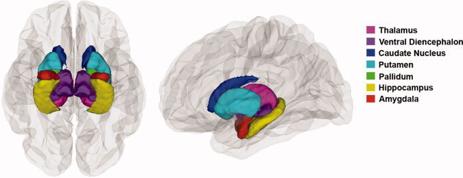
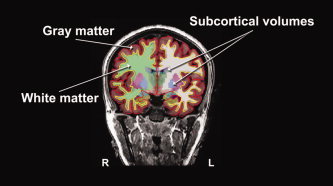
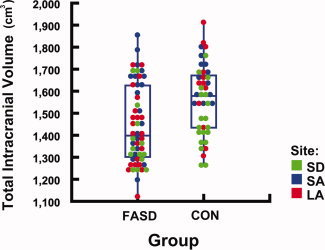
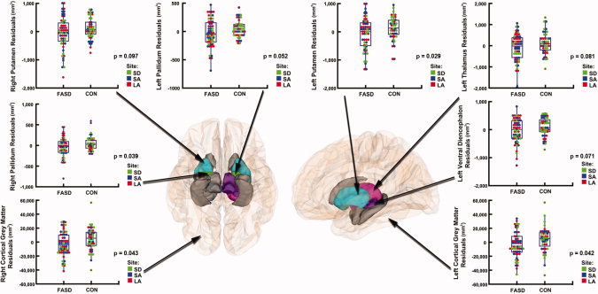
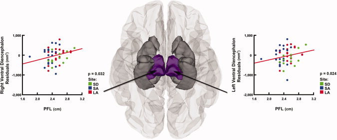
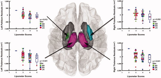
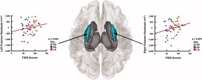
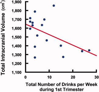
Individuals with heavy prenatal alcohol exposure can experience significant deficits in cognitive and psychosocial functioning and alterations in brain structure that persist into adulthood. In this report, data from 99 participants collected across three sites (Los Angeles and San Diego, California, and Cape Town, South Africa) were analyzed to examine relationships between brain structure, neurocognitive function, facial morphology, and maternal reports of quantities of alcohol consumption during the first trimester. Across study sites, we found highly significant volume reductions in the FASD group for all of the brain regions evaluated. After correcting for scan location, age, and total brain volume, these differences remained significant in some regions of the basal ganglia and diencephalon. In alcohol-exposed subjects, we found that smaller palpebral fissures were significantly associated with reduced volumes in the ventral diencephalon bilaterally, that greater dysmorphology of the philtrum predicted smaller volumes in basal ganglia and diencephalic structures, and that lower IQ scores were associated with both smaller basal ganglia volumes and greater facial dysmorphology. In subjects from South Africa, we found a significant negative correlation between intracranial volume and total number of drinks per week in the first trimester. These results corroborate previous reports that prenatal alcohol exposure is particularly toxic to basal ganglia and diencephalic structures. We extend previous findings by illustrating relationships between specific measures of facial dysmorphology and the volumes of particular subcortical structures, and for the first time show that continuous measures of maternal alcohol consumption during the first trimester relates to overall brain volume reduction.
Copyright © 2011 Wiley Periodicals, Inc.
Figures
Similar articles
-
Callosal thickness reductions relate to facial dysmorphology in fetal alcohol spectrum disorders.Alcohol Clin Exp Res. 2012 May;36(5):798-806. doi: 10.1111/j.1530-0277.2011.01679.x. Epub 2011 Dec 7. Alcohol Clin Exp Res. 2012. PMID: 22150665 Free PMC article.
-
Abnormal cortical thickness alterations in fetal alcohol spectrum disorders and their relationships with facial dysmorphology.Cereb Cortex. 2012 May;22(5):1170-9. doi: 10.1093/cercor/bhr193. Epub 2011 Jul 28. Cereb Cortex. 2012. PMID: 21799209 Free PMC article.
-
A longitudinal study of the long-term consequences of drinking during pregnancy: heavy in utero alcohol exposure disrupts the normal processes of brain development.J Neurosci. 2012 Oct 31;32(44):15243-51. doi: 10.1523/JNEUROSCI.1161-12.2012. J Neurosci. 2012. PMID: 23115162 Free PMC article.
-
Neuroimaging effects of prenatal alcohol exposure on the developing human brain: a magnetic resonance imaging review.Acta Neuropsychiatr. 2015 Oct;27(5):251-69. doi: 10.1017/neu.2015.12. Epub 2015 Mar 17. Acta Neuropsychiatr. 2015. PMID: 25780875 Review.
-
Imaging the impact of prenatal alcohol exposure on the structure of the developing human brain.Neuropsychol Rev. 2011 Jun;21(2):102-18. doi: 10.1007/s11065-011-9163-0. Epub 2011 Mar 3. Neuropsychol Rev. 2011. PMID: 21369875 Free PMC article. Review.
Cited by
-
White matter deficits mediate effects of prenatal alcohol exposure on cognitive development in childhood.Hum Brain Mapp. 2016 Aug;37(8):2943-58. doi: 10.1002/hbm.23218. Epub 2016 May 24. Hum Brain Mapp. 2016. PMID: 27219850 Free PMC article.
-
Sexual dimorphism of volume reduction but not cognitive deficit in fetal alcohol spectrum disorders: A combined diffusion tensor imaging, cortical thickness and brain volume study.Neuroimage Clin. 2017 May 10;15:284-297. doi: 10.1016/j.nicl.2017.05.006. eCollection 2017. Neuroimage Clin. 2017. PMID: 28560153 Free PMC article.
-
White matter microstructure in fetal alcohol spectrum disorders: A systematic review of diffusion tensor imaging studies.Hum Brain Mapp. 2019 Feb 15;40(3):1017-1036. doi: 10.1002/hbm.24409. Epub 2018 Oct 5. Hum Brain Mapp. 2019. PMID: 30289588 Free PMC article.
-
Qualitative and Quantitative Comparison of Hippocampal Volumetric Software Applications: Do All Roads Lead to Rome?Biomedicines. 2022 Feb 12;10(2):432. doi: 10.3390/biomedicines10020432. Biomedicines. 2022. PMID: 35203641 Free PMC article.
-
Atypical fetal development: Fetal alcohol syndrome, nutritional deprivation, teratogens, and risk for neurodevelopmental disorders and psychopathology.Dev Psychopathol. 2018 Aug;30(3):1063-1086. doi: 10.1017/S0954579418000500. Dev Psychopathol. 2018. PMID: 30068419 Free PMC article. Review.
References
-
- Adnams CM, Kodituwakku PW, Hay A, Molteno CD, Viljoen D, May PA ( 2001): Patterns of cognitive‐motor development in children with fetal alcohol syndrome from a community in South Africa. Alcohol Clin Exp Res 25: 557–562. - PubMed
-
- Archibald SL, Fennema‐Notestine C, Gamst A, Riley EP, Mattson SN, Jernigan TL ( 2001): Brain dysmorphology in individuals with severe prenatal alcohol exposure. Dev Med Child Neurol 43: 148–154. - PubMed
Publication types
MeSH terms
Grants and funding
- U24AA014811/AA/NIAAA NIH HHS/United States
- U01 AA011685/AA/NIAAA NIH HHS/United States
- R01 AA015134/AA/NIAAA NIH HHS/United States
- U24 AA014811/AA/NIAAA NIH HHS/United States
- P41 RR013642/RR/NCRR NIH HHS/United States
- R01 HD053893-01/HD/NICHD NIH HHS/United States
- R01 DA017830/DA/NIDA NIH HHS/United States
- U01 AA017122/AA/NIAAA NIH HHS/United States
- R01 HD053893/HD/NICHD NIH HHS/United States
- U01 AA014834/AA/NIAAA NIH HHS/United States
- UO1 AA 11685/AA/NIAAA NIH HHS/United States
- U54 RR021813/RR/NCRR NIH HHS/United States
- R01 AA15134/AA/NIAAA NIH HHS/United States
- R01 DA017831/DA/NIDA NIH HHS/United States
- U01 AA017122-01/AA/NIAAA NIH HHS/United States
LinkOut - more resources
Full Text Sources
Medical
