. 2011 Jun 17;6(6):636-47.
doi: 10.1021/cb2000346. Epub 2011 Apr 20.

Small molecule inhibition of the TNF family cytokine CD40 ligand through a subunit fracture mechanism

Affiliations

Small molecule inhibition of the TNF family cytokine CD40 ligand through a subunit fracture mechanism

Laura F Silvian et al. ACS Chem Biol. .

Erratum in

  • ACS Chem Biol. 2011 Jul 15;6(7):761. Shipps, Gerald W [added]

Abstract

BIO8898 is one of several synthetic organic molecules that have recently been reported to inhibit receptor binding and function of the constitutively trimeric tumor necrosis factor (TNF) family cytokine CD40 ligand (CD40L, aka CD154). Small molecule inhibitors of protein-protein interfaces are relatively rare, and their discovery is often very challenging. Therefore, to understand how BIO8898 achieves this feat, we characterized its mechanism of action using biochemical assays and X-ray crystallography. BIO8898 inhibited soluble CD40L binding to CD40-Ig with a potency of IC(50) = 25 μM and inhibited CD40L-dependent apoptosis in a cellular assay. A co-crystal structure of BIO8898 with CD40L revealed that one inhibitor molecule binds per protein trimer. Surprisingly, the compound binds not at the surface of the protein but by intercalating deeply between two subunits of the homotrimeric cytokine, disrupting a constitutive protein-protein interface and breaking the protein's 3-fold symmetry. The compound forms several hydrogen bonds with the protein, within an otherwise hydrophobic binding pocket. In addition to the translational splitting of the trimer, binding of BIO8898 was accompanied by additional local and longer-range conformational perturbations of the protein, both in the core and in a surface loop. Binding of BIO8898 is reversible, and the resulting complex is stable and does not lead to detectable dissociation of the protein trimer. Our results suggest that a set of core aromatic residues that are conserved across a subset of TNF family cytokines might represent a generic hot-spot for the induced-fit binding of trimer-disrupting small molecules.


Figures
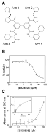
Figure 1
Figure 1
Structure and activity of BIO8898. a) Chemical structure of BIO8898. b) BIO8898 inhibits the binding of 40 ng/ml mycCD40L to CD40-Ig with IC50 ~25 μM. CD40-Ig was coated on the assay plate, and the binding of mycCD40L was measured after one hour by DELFIA using a europium-labeled anti-myc antibody, as described in Materials and Methods. Data points are the average of triplicate measurements from a single experiment, with the standard deviation shown by the error bars. The solid line represents the best fit of a hyperbolic (single site) binding equation. c) BIO8898 inhibits the ability of 3 ng/ml mycCD40L plus 50 μg/ml cyclohexamide to induce apoptosis in BHK cells transfected with a chimeric receptor comprising the extracellular domain of CD40 fused to the transmembrane and cytoplasmic domains of the structurally related TNF family receptor TNFR2, or p75. The number of viable cells present after two days was determined by MTT staining, as described in Materials and Methods. The absorbance of control wells containing the same concentration of DMSO but no BIO8898 was subtracted from each measurement. A higher absorbance at 590 nm indicates more live cells. Data are plotted as the average of two independent experiments, with the error bars showing the spread between the values observed in each experiment. Inset plot shows the same data plotted on a linear rather than a logarithmic concentration axis. The BIO8898 dose-response curve does not reach saturation because compound concentrations were limited by poor solubility at the low DMSO levels that the assay can tolerate. The solid line represents the best fit to a hyperbolic binding equation (EC50 = 69 μM).
Figure 2
Figure 2
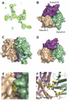
Structure of BIO8898/CD40L complex. a) Sigma-A weighted omit map density contoured around BIO8898 at 1 sigma. b) Space-filling representation of native CD40L trimer, as viewed from the top in relation to the three-fold axis of symmetry, to compare with BIO8898-bound CD40L: Subunit A (green), Subunit B (magenta), subunit C (tan). c) Side view and (D) top view of the complex of BIO8898 with CD40L. The atoms of the inhibitor are colored as follows: C, yellow; N, blue; O, red. The atoms of the CD40L glycan that were visible in the structure are shown in stick representation (dark blue) and the subunits are colored according to b). e) Closer view of the inhibitor binding region from (C) with protein subunits A and C rendered partially transparent to better show the orientation of the bound compound and the extent to which it is buried within the protein. f) Closer view of the inhibitor binding region, with CD40L subunits shown in ribbon representation to illustrate the binding locations of the four arms of the bound BIO8898.
Figure 3
Figure 3
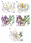
Details of the intermolecular interactions between BIO8898 and CD40L. a) Hydrogen bonding interactions connecting protein, ligand, and ordered water molecules in the binding site. b) Ribbons representation of the rigid-body conformational changes of CD40L bound to BIO8898 (colored by subunits) when superimposed with subunit B of unbound CD40L (grey). c) Residues Tyr170 and His224 (shown in space-filling representation) contributed by each of the three CD40L subunits form a compact core in the native trimer. Subunit A has been removed from the representation, except for residues Y170 and H224 (shown in pale brown), to allow a clearer view of the core interactions. d) Same view as in c) but for the BIO8898/CD40L complex showing how Tyr170 and His224 conformationally adapt to accommodate the inhibitor. e) The AA” loop of subunit A undergoes a substantial conformational change from its position in the native trimer (shown in grey) to the position it occupies in the complex with BIO8898 (shown in pale brown) in which it covers the compound (yellow stick).
Figure 4
Figure 4
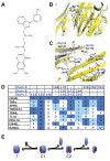
Comparison of BIO8898/CD40L complex to the SP307/TNFα complex reported in He et al. a) Structure of inhibitor SP307 (12). b) Superposition of backbone C-alphas of SP307/TNFalpha complex (grey) and the BIO8898/CD40L (yellow), with CD40L subunit A removed for clarity, illustrating that the SP307 and Arm 4 of BIO8898 bind in corresponding locations in the core of the protein. The N-termini of the protein are at the bottom of the figure and is thus reversed from the orientation shown in Figure 3.c) Similar side-chains are involved in binding SP307 to TNFα (grey with black labels) and BIO8898 to CD40L (yellow with blue labels), including many conserved aromatic residues. d) Structure-based sequence alignment of CD40L with other TNF-family members illustrating conservation in residues contacting BIO8898. In particular, residues 170, 172 and 224, which are exploited for BIO8898 interactions in more than 1 subunit, and are highly homologous across several family TNF members. Color coding is as follows: dark blue, exact identity with CD40L; light blue, similar aromatic or hydrophobic side-chains to those in CD40L. e) Mechanism proposed by He et al., for the inhibition of TNFα by SP307, in which T represents the intact trimeric cytokine and C2 represents the complex of inhibitor with dimeric cytokine that was observed for TNFα with SP307. The species C1 corresponds to a complex in which the inhibitor is intercalated into the protein trimer, as we report here for CD40L with BIO8898.
Figure 5
Figure 5
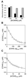
Dissociation and stoichiometry assays. a) BIO8898 dose-dependently inhibits the binding of CD40-Ig to sparsely biotinylated CD40L that has been captured via a single biotin per trimer on a neutravidin-coated assay plate (black bars). However, if the assay plate is washed prior to CD40-Ig addition, to remove compound plus any dissociated CD40L subunits, no inhibition is observed (grey bars), showing that binding of BIO8898 does not cause irreversible disruption of CD40L or full ejection of CD40L subunits from the complex. The results shown are from a single experiment. b) Binding of mycCD40L to CD40-Ig in solution, as measured using the initial rates Biacore titration method. Pre-incubation of individual samples each containing 100 nM CD40L with the indicated concentrations of CD40-Ig results in detection of lower concentrations of free CD40L, as the CD40L molecules that are bound by CD40-Ig in solution at equilibrium are blocked from binding to the CD40-Ig on the Biacore chip and thus are not detected. The solid line represents the best fit of the CD40L/CD40-Ig data to a quadratic binding equation, giving an apparent KD for this interaction of 7.6 ± 2.1 nM (n = 13). c) Similar experiment to that shown in (b), except that samples containing fixed concentrations of 1.5 M CD40L were pre-incubated with 0-24 M monomeric sCD40 before each mixture was analyzed by Biacore. The data represent the mean of two Biacore measurements of each solution, separated by several hours. Error bars showing the spread of the duplicate data points are included, but in most cases are smaller than the data symbol so cannot be seen. The solid line represents the best fit to a hyperbolic binding equation, indicating that CD40L binds monomeric sCD40 in solution only weakly, with KD = 13 ± 2 M (n = 2).

References

    1. Wells JA, McClendon CL. Reaching for high-hanging fruit in drug discovery at protein-protein interfaces. Nature. 2007;450:1001–1009. - PubMed
    1. Gerrard JA, Hutton CA, Perugini MA. Inhibiting protein-protein interactions as an emerging paradigm for drug discovery. Mini Rev Med Chem. 2007;7:151–157. - PubMed
    1. Fischer P. Protein-Protein Interactions in Drug Discovery. Drug Design Reviews -Online. 2005;2:179–207.
    1. Whitty A, Kumaravel G. Between a rock and a hard place? Nat Chem Biol. 2006;2:112–118. - PubMed
    1. Ganesan A. The impact of natural products upon modern drug discovery. Curr Opin Chem Biol. 2008;12:306–317. - PubMed

Publication types

MeSH terms

Substances